
Posterior tibial tendon dysfunction exercises and physiotherapy
What is posterior tibial tendon dysfunction?
The tibialis posterior is a deep muscle at the back of your lower leg. Its tendon wraps around the back of the inner ankle bone and attaches under the inner arch, and it helps hold the arch up when you stand and walk. In posterior tibial tendon dysfunction (PTTD), that tendon becomes painful and weak, and over time it can stretch or tear. The arch then sags and the heel bone tips outward, which puts more strain on the ligaments and joints around it.
OrthoInfo, from the American Academy of Orthopaedic Surgeons, now calls it progressive collapsing foot deformity. Women and people over 40 get it more often. OrthoInfo lists obesity and diabetes as risk factors, and high blood pressure too. It can follow an injury such as a fall, or build up from overuse.
Typical signs are pain, sometimes with swelling, along the inside of the foot and ankle, weakness when you push off, and trouble walking or standing for long. As the heel shifts outward, the outside of the ankle can start to hurt as well.
Two simple checks help. Stand on the sore leg alone and try to rise onto tiptoe: according to OrthoInfo, you need a healthy tibialis posterior tendon to do this, so difficulty points to a tendon problem. Seen from behind, a collapsing foot shows more of the little toes on the outside than the other foot does, which is called the "too many toes" sign. A doctor or physio will check both and compare sides.
The stages of posterior tibial tendon dysfunction
The classic staging comes from Johnson and Strom in 1989, later extended by Myerson. It follows the tendon as it fails (Pasapula and colleagues, 2024). In stage 1 the tendon is sore and inflamed, but the arch holds and the foot keeps its shape. In stage 2 the tendon has stretched or partly torn and the arch has dropped, but the foot is still flexible, so it can be moved back into shape, and rising onto tiptoe on that leg alone is usually hard. In stage 3 the collapse has become stiff (fixed), and stage 4 brings in the ankle joint too, as the strong ligament on the inside of the ankle (deltoid ligament) gives way.
In 2020 an expert group proposed the name progressive collapsing foot deformity and a new system (Myerson and colleagues, 2020). It grades the foot as flexible (stage 1) or rigid (stage 2), then describes where in the foot the collapse is (Pasapula and colleagues, 2024). For you, the main question is the same in both systems: is the foot still flexible, or has it become stiff? The exercise research on this page was done in people with early, flexible stages (Ross and colleagues, 2018).
How this differs from flat feet
Many adults have flat feet all their lives and never have pain. The flat feet program covers those feet and painful flexible flat feet in general, including what insoles can do. This page is for the tendon problem: an arch that used to be normal and is now dropping, or pain and weakness along the tibialis posterior tendon. The two programs share several exercises, but this one spends more time on the tendon itself and on braces, and explains when surgery comes in.
Why does exercise help posterior tibial tendon dysfunction?
A weak tendon needs to take load again, and the research, though small, supports strengthening. In a trial of 36 adults with stage 1 or 2 tendon problems, every group wore orthoses and did calf stretches for 12 weeks (Kulig and colleagues, 2009). One group added exercise that shortened the muscle against resistance (concentric), and another added slow lowering (eccentric). All three groups had less pain and better foot function, with the biggest gains in the eccentric group and the smallest with orthoses and stretching only. Strengthening without the slow lowering did not clearly beat orthoses and stretching alone, so the support and stretching may do part of the work (Ross and colleagues, 2018).
A 2018 systematic review found only 3 randomized trials, with 93 people in total, all in early stages (Ross and colleagues, 2018). Adding eccentric strengthening to stretching and orthoses had a moderate effect on pain and disability. It also found that the trials described their exercise doses poorly, and it concluded that high-quality research is scarce. So strengthening is a sensible first step with some support behind it, not a proven cure.
A clinic series points the same way. In 47 people with stage 1 or 2 tendon problems, treatment combined a short hinged ankle-foot brace (orthosis) or a foot orthosis with high repetition strengthening and a home program that included calf stretches (Alvarez and colleagues, 2006). After a median of 10 visits to a physical therapist (physiotherapist) over a median of 4 months, 39 of the 47 had a good result and 5 went on to surgery. There was no comparison group, so the study cannot show how much came from the brace and how much from the exercise.
Insoles, braces and boots
Support takes load off the tendon while it gets stronger. OrthoInfo suggests ready-made orthotics for a mild deformity and custom-made ones for moderate to severe cases. For braces, it suggests a ready-made lace-up ankle brace for mild to moderate flatfoot and a heavier-duty brace for severe cases. In the Kulig and Alvarez studies, everyone wore an orthosis or brace, so exercise was always added to support rather than used instead of it.
OrthoInfo describes a short leg cast or walking boot for 6 to 8 weeks to rest the tendon and let the swelling go down. Because a cast weakens the other leg muscles, it is mainly used when other treatment has not worked, and a brace usually follows. It also lists cutting back on the activities that make the pain worse, cold packs for 20 minutes 3 to 4 times a day, and anti-inflammatory painkillers. Check with a pharmacist before taking them if you are unsure they are safe for you. Cycling or swimming in place of some walking and standing spares the foot while the tendon settles, and OrthoInfo notes that losing weight, if you are overweight, takes stress off the arch.
How to use this program
The three phases below are steps in the exercise program, not the medical stages above. The program is meant for a foot that is still flexible: stage 1 or 2 in the older system, or stage 1 in the 2020 system. If your foot has become stiff, or your doctor has told you it is stage 3 or 4, or stage 2 (rigid) in the 2020 system, ask your physio or surgeon which parts suit you before you start. Start at the phase that fits how your foot is now. When in doubt, begin with phase 1 and step up once it feels easy and the next morning is no worse.
Phase 1 should not hurt at all. From phase 2 on, a little discomfort while you exercise is usually fine, as long as it fades soon afterward and the foot is no worse the next morning. A clearly sorer inner ankle or arch the next day means you did too much. Go back a step instead of stopping. Keep calf stretches short of any pain on the inside of the ankle: a stretch at full range both squeezes and pulls on the tendon where it curves behind the ankle bone, and that mix may irritate it rather than help (Ross and colleagues, 2018).
In the two 12 week trials, people did their tendon exercises twice a day, in the main trial as 3 sets of 15 against a load they could only just manage 15 times (Ross and colleagues, 2018). The exercise pages in this program start lower, mostly at 2 to 3 sets of 10 to 15, and each one gives a typical starting dose. Your physio will adjust the dose to your foot and to the activities you want to return to. Expect slow change: OrthoInfo warns the pain may last longer than 3 months even with early treatment. Compare your foot from week to week, not day to day.
Do the standing and balance work next to a kitchen counter or a wall, and hold on if you feel unsteady. If you have diabetes or numb feet, look over the skin of both feet once each session is done. Ask your doctor, podiatrist or physio first about any exercise that presses or rubs on the sole.
The exercise program
Phase 1: Settle the tendon and start loading it gently
For the first weeks, or while the inside of your ankle aches with ordinary walking and standing. If you have been given an insole or brace, wear it for daily walking as advised. The isometric inversion works the tibialis posterior with no movement at the ankle, and the towel sweeps add gentle movement with the foot supported on the floor. The seated calf raise builds calf strength without your full body weight on the foot. None of these should hurt, and the calf stretches should stay gentle: stop before your deepest stretch and short of any pain on the inside of the ankle, as a full stretch squeezes the tendon there, and do not let the arch roll in.
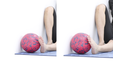 Isometric ankle inversion
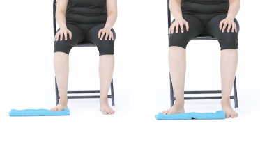 Foot towel sweeps
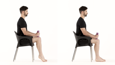 Seated calf raise
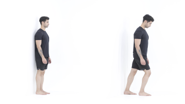 Calf stretch
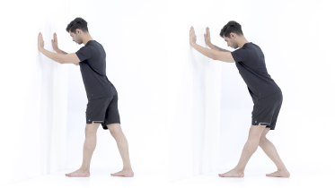 Soleus stretch
Phase 2: Strengthen the tendon against resistance
When everyday walking is comfortable and phase 1 no longer brings on pain. The band exercise is similar to the one used in the main trial, which turned the foot inward against resistance, though in the trial the foot was pointed down (Kulig and colleagues, 2009). Let the foot return slowly each time, because in that trial the people who trained the slow return (eccentric) did best. After the band, do calf raises on both feet, and add the toes-in version once plain raises feel easy. Balance on the foam pad trains the small ankle corrections you need on uneven ground.
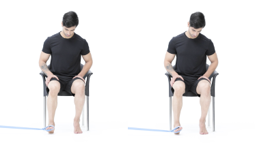 Ankle inversion with band
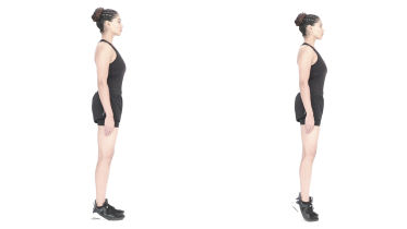 Calf raises
 Calf raise with toes turned in
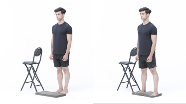 Standing balance on a foam pad
Phase 3: Single leg heel raises and longer walks
Move on once you can do 2 to 3 sets of double leg calf raises easily and the foot feels no worse the following morning. The eccentric calf raise is the stepping stone: rise with both feet, then lower slowly on the painful side alone. Rising onto tiptoe on one leg is the test doctors use for this tendon, so single leg heel raises make a good goal, with your fingertips on a counter for balance. Toe walking and single leg balance prepare the foot for longer walks. In one clinic program, people worked toward 50 single leg heel raises and 100 feet (about 30 m) of toe walking with little or no pain (Alvarez and colleagues, 2006).
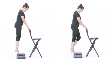 Eccentric calf raise
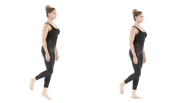 Single leg heel raise
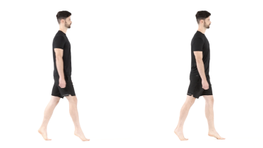 Toe walking
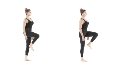 Single leg stance
When is surgery considered?
OrthoInfo's advice is to consider surgery when several months of appropriate treatment have not eased the pain. The choice of operation depends on how severe the deformity is, whether there is arthritis and how flexible the foot still is. Operations range from cleaning up the inflamed tendon, lengthening a tight calf, moving a nearby toe tendon across to take over from the damaged one, repairing ligaments and reshaping the heel bone, to fusing joints, which is more common when the foot is stiff or arthritic. Severe cases can need surgery on the ankle as well.
OrthoInfo says most people have good results from surgery, but healing takes 6 months or more. After an operation on your foot or ankle, follow the rehab plan your surgeon gives you instead of this page.
When to see a physio or doctor
Get your foot checked by a physio or doctor if pain along the inside of your ankle is not settling, if your arch looks lower than it used to or lower than on the other foot, or if rising onto tiptoe on one leg has become hard. A physio or podiatrist can check the tendon and advise on insoles or a brace. They can also tell you which phase to start at. If your foot is not improving after several months of steady treatment, ask to see a foot and ankle specialist. The warning signs listed further down need prompt medical attention.
This program is meant for adults. If a child's flat feet are painful or stiff, or only one foot is flat, have a doctor or physio check them.
For physiotherapists
The program here is a patient starting point for stage 1 and 2 PTTD, which the 2020 consensus calls flexible progressive collapsing foot deformity (Pasapula and colleagues, 2024). Compare the single limb heel rise and the too many toes sign with the other side, and check whether the hindfoot corrects non-weight-bearing. Refer a rigid deformity, or ankle valgus from deltoid failure, for an orthopedic opinion, since loading alone is not enough there. Before loading anyone with diabetes or neuropathy, exclude active Charcot neuroarthropathy (the patient warning signs ask for same-day advice). Ask about recent fluoroquinolone antibiotics (MHRA, 2024).
Kulig and colleagues (2009) randomized 36 adults to 12 weeks of orthoses and stretching, alone or with concentric or eccentric resisted foot adduction in plantarflexion. As reported by Ross and colleagues (2018), the dose was 3 sets of 15 at a 15 repetition maximum, twice a day, with about 5 seconds under tension per repetition. The other two trials used 2 to 3 sets of 10 to 30 twice daily for 12 weeks (heel raises, banded adduction and inversion in plantarflexion, single leg heel raises) and 3 to 4 sets of 15 to 20 over 6 weeks. Ross and colleagues reported moderate effects (SMD 0.6 to 1.2) when eccentric work was added to stretching and orthoses, but concluded that no firm dosing recommendation can be made.
Ross and colleagues also note that static calf stretching to end-range dorsiflexion loads the tendon behind the medial malleolus in compression as well as tension. Cue patients to stop stretches before medial ankle pain. Alvarez and colleagues (2006) used a short articulated AFO or foot orthosis with high repetition strengthening, and discharged patients at no more than a 10% strength deficit, 50 single support heel rises and 100 feet of toe walking with minimal or no pain, and 200 repetitions of each home exercise. These make practical targets, though they come from an uncontrolled series. Let the 24-hour response, not a set schedule, decide when to progress.
See a doctor promptly if
- You have diabetes or numb feet, and one foot becomes hot, swollen or red, or changes color or shape, with or without pain. Take your weight off that foot straight away and get advice the same day from your doctor or your diabetes foot team. This can be Charcot foot, where the bones and joints of the foot are damaged and the foot can collapse. Do not exercise that foot until it has been checked. If you also have a fever or feel very unwell, or redness is spreading up your foot or leg, go to an emergency department.
- The foot is hot, red and swollen, or you have a fever or feel unwell. Get medical help the same day, and go to an emergency department if you feel very unwell. If you have a very high or very low temperature, shivering you cannot control, you are breathing very fast or finding it hard to breathe, you are confused or your speech is slurred, your skin, lips or tongue look blue, gray, pale or blotchy (on brown or black skin this can be easier to see on the palms of the hands or soles of the feet), or you have a rash that does not fade when you press it, call emergency services, as this can be sepsis.
- You have diabetes or numb feet and find a new blister, cut or sore on your foot, especially one you did not feel, a wound that smells or is leaking, or new pain in your foot that you cannot explain. Get it seen by your doctor or foot team the same day. If you also have a fever or feel very unwell, redness is spreading up your foot or leg, or part of your foot or a toe turns black, go to an emergency department.
- Part of your foot or a toe turns cold, pale, blue or black. Call emergency services or go to an emergency department straight away, and do not drive yourself, as the blood supply to the foot may be blocked.
- You felt a sudden pop or snap on the inside of your ankle, or a sudden sharp pain there, and since then you can no longer rise onto tiptoe on that leg when you could before, or the arch has suddenly dropped. Stop the exercises, take the weight off that foot as much as you can and get it checked the same day at an urgent care center or emergency department, even if you can still walk. The tendon that holds up the arch (tibialis posterior) may have torn.
- Your tendon pain or swelling started while you were taking a fluoroquinolone antibiotic, such as ciprofloxacin or levofloxacin, or in the months after a course. Contact your doctor straight away. The UK medicines regulator (MHRA) advises stopping the antibiotic at the first sign of tendon pain or swelling and talking to your doctor. Hold off the foot and ankle exercises until you have been checked.
- The pain started with a fall, a twist or a sudden pop on the inside of the ankle, and there is swelling or bruising, or you cannot put weight on the foot. Get it checked the same day.
- A calf or thigh that is newly swollen, warm, tender, red or darker than usual, has swollen veins that are sore to touch, or has a throbbing or cramping pain that does not fit your injury or condition, or that feels different from normal muscle soreness after exercise. This can be a sign of a blood clot (deep vein thrombosis), especially after surgery, an injury, time in a cast or boot, a long trip or a spell of being much less mobile than usual. Get medical advice the same day. If you are also short of breath or have chest pain, call emergency services.
- The arch on one foot has dropped, suddenly or over weeks or months, or you can no longer rise onto tiptoe standing on that leg alone, especially with pain or swelling along the inside of the ankle. See a doctor within a few days. This can be a sign that the tendon that holds up the arch (tibialis posterior) is failing, and it can keep getting worse without treatment. If you have diabetes or numb feet, a change in the shape of your foot is a same-day sign, as described above.
- The pain is there at night or at rest as well as when you stand and walk, and it is getting worse. See your doctor within a few days. If you have had cancer, now or in the past, or are losing weight without trying, see your doctor sooner, within a day or two, and mention it. This applies even if the pain is not getting worse. If you are being treated for cancer now, contact your cancer team the same day.
- You have new numbness, tingling, burning or loss of feeling in the foot or toes. Book an assessment with a doctor or physio in the next week or two.
Common questions
Can posterior tibial tendon dysfunction heal without surgery?
Often it can, especially when it is treated while the foot is still flexible. In a clinic series of 47 people with stage 1 or 2 tendon problems, 39 (83%) had a good result with a brace or orthosis and a structured exercise program, and 5 (11%) needed surgery (Alvarez and colleagues, 2006). That study had no comparison group, and the research is on early stages, so it says less about a foot that has already become stiff. Your physio will adjust the program to your foot, and a foot and ankle specialist can advise if it is not improving.
How long does posterior tibial tendon dysfunction take to heal?
Usually months. Even with early treatment, OrthoInfo says the pain may go on for longer than 3 months. The exercise trials ran for 12 weeks, and in the clinic series by Alvarez and colleagues (2006), treatment took a median of 4 months. After surgery, OrthoInfo says healing takes 6 months or more.
Do I need a brace for posterior tibial tendon dysfunction?
In the Kulig and Alvarez studies, everyone wore an orthosis or brace alongside their exercises. OrthoInfo suggests a ready-made ankle brace for mild to moderate flatfoot and a heavier-duty brace for severe cases, with ready-made or custom orthotics as the other option, depending on how severe the deformity is. A walking boot or short leg cast for 6 to 8 weeks is sometimes used to rest the tendon, mostly when other treatment has not worked, with a brace to follow once the pain and swelling settle. A physio, podiatrist or orthotist can advise what suits your foot.
Is walking good for posterior tibial tendon dysfunction?
Walking is usually fine at a level your tendon tolerates, but long walks and long spells of standing are often what hurts. OrthoInfo advises cutting back on activities that make the pain worse and swapping some of them for lower impact exercise, such as cycling or swimming, while the tendon settles. If you have been given an insole or brace, wear it for walking. If the inside of your ankle is clearly worse the next morning after a walk, make the next one shorter.
Can a steroid injection help posterior tibial tendon dysfunction?
OrthoInfo says cortisone injections are not normally given into the posterior tibial tendon, because of the risk that the tendon tears. Whether any injection is worth it is a question for your doctor. Support and strengthening stay the main treatment without surgery.
References
- American Academy of Orthopaedic Surgeons. Progressive Collapsing Foot Deformity (Flatfoot). OrthoInfo. https://www.orthoinfo.org/en/diseases--conditions/posterior-tibial-tendon-dysfunction/
- Kulig K, Reischl SF, Pomrantz AB, Burnfield JM, Mais-Requejo S, Thordarson DB, Smith RW. Nonsurgical management of posterior tibial tendon dysfunction with orthoses and resistive exercise: a randomized controlled trial. Physical Therapy. 2009;89(1):26-37. https://doi.org/10.2522/ptj.20070242
- Ross MH, Smith MD, Mellor R, Vicenzino B. Exercise for posterior tibial tendon dysfunction: a systematic review of randomised clinical trials and clinical guidelines. BMJ Open Sport and Exercise Medicine. 2018;4(1):e000430. https://doi.org/10.1136/bmjsem-2018-000430
- Alvarez RG, Marini A, Schmitt C, Saltzman CL. Stage I and II posterior tibial tendon dysfunction treated by a structured nonoperative management protocol: an orthosis and exercise program. Foot and Ankle International. 2006;27(1):2-8. https://doi.org/10.1177/107110070602700102
- Myerson MS, Thordarson DB, Johnson JE, Hintermann B, Sangeorzan BJ, Deland JT, Schon LC, Ellis SJ, de Cesar Netto C. Classification and nomenclature: progressive collapsing foot deformity. Foot and Ankle International. 2020;41(10):1271-1276. https://doi.org/10.1177/1071100720950722
- Pasapula CS, Choudkhuri MR, Monzó ERG, Dhukaram V, Shariff S, Pasterse V, Richie D, Kobezda T, Solomou G, Cutts S. Review of classification systems for adult acquired flatfoot deformity/progressive collapsing foot deformity and the novel development of the triple classification delinking instability/deformity/reactivity and foot type. Journal of Clinical Medicine. 2024;13(4):942. https://doi.org/10.3390/jcm13040942
- Medicines and Healthcare products Regulatory Agency. Fluoroquinolone antibiotics: must now only be prescribed when other commonly recommended antibiotics are inappropriate. Drug Safety Update. 22 January 2024. https://www.gov.uk/drug-safety-update/fluoroquinolone-antibiotics-must-now-only-be-prescribed-when-other-commonly-recommended-antibiotics-are-inappropriate
- Diabetes UK. Charcot foot and diabetes. Last reviewed 8 November 2024. https://www.diabetes.org.uk/about-diabetes/looking-after-diabetes/complications/feet/charcot-foot
- Diabetes UK. Serious foot problems and diabetes. Last reviewed 24 December 2024. https://www.diabetes.org.uk/about-diabetes/looking-after-diabetes/complications/feet/serious-foot-problem
- National Institute for Health and Care Excellence. Diabetic foot problems: prevention and management. NICE guideline NG19. Published 26 August 2015, last updated 11 October 2019. https://www.nice.org.uk/guidance/ng19
- NHS. Sepsis. Page last reviewed 14 May 2026. https://www.nhs.uk/conditions/sepsis/
- NHS. Deep vein thrombosis (DVT). Page last reviewed 30 April 2026. https://www.nhs.uk/conditions/deep-vein-thrombosis-dvt/
Written and checked by the PocketPhysio editorial team. Last updated 2026-09-28.
This page is general education, not a diagnosis or a personal treatment plan. See a physiotherapist or doctor for advice about your own situation.