
Peroneal tendinopathy exercises and physiotherapy
What is peroneal tendinopathy?
The peroneal muscles, also called fibularis longus and brevis, run down the outside of your lower leg. Their tendons curve behind the bony bump on the outside of your ankle, where a band of tissue holds them in a groove, then run along the outer side of the heel into the foot. They turn the foot outward and help steady the ankle, especially on uneven ground. Peroneal tendinopathy is pain in these tendons that comes with loading them.
The usual story is pain and sometimes swelling behind the outer ankle bone or along the outside of the heel. The EFORT review describes pain that gets worse with activity and better with rest, tenderness along the tendons, and pain when you push the foot outward against resistance or when it is turned inward on a stretch (Davda and colleagues, 2017). It is often worse on slopes or rough ground, and when you turn.
Two things often come before it. One is an ankle sprain, or an ankle that keeps giving way afterward (Davda and colleagues, 2017; van Dijk and colleagues, 2018). The other is foot shape: a high arch with a heel that tips inward (cavovarus) puts more load through these tendons, and the European consensus names it as a factor that may lead to tendon problems.
This program is written for adults. A child or teenager with ankle pain should be checked by a doctor or physio before starting any program. If you have had surgery on these tendons, follow your surgeon's program rather than this page.
How it differs from an ankle sprain
A lateral ankle sprain is a sudden injury to the ligaments in front of and below the outer ankle bone, usually from rolling the foot inward. The ankle sprain program covers that injury, including when you need an X-ray. Peroneal tendinopathy sits just behind the bone, usually builds up over weeks, and hurts most when the tendons work.
The two overlap. A sprain can injure these tendons at the same time, and the US physical therapy guideline for ankle sprains lists injury to the fibularis tendons among the problems to consider when an ankle does not recover as expected (Martin and colleagues, 2021). If the main problem is an ankle that keeps giving way, not an ache behind the bone, start with the balance and bracing advice on the ankle sprain page. A physio can check which one you have.
Why does exercise help peroneal tendinopathy?
Tendons respond to load, and a tendon that is rested for a long time tends to lose strength. The European consensus advises that treatment without surgery should be considered for everyone with a peroneal tendon problem. After a new injury, it describes starting to walk on the foot once that is pain free, then range of motion and exercises that load the tendon (van Dijk and colleagues, 2018). Where it describes rehab without surgery, for tendons that have slipped out of place, the exercises are peroneal strengthening and balance (proprioception) training.
The evidence here is thin. There are no good trials of exercise programs for peroneal tendinopathy itself, and the EFORT review notes that treatment advice for these tendons rests mainly on case series and expert opinion (Davda and colleagues, 2017). The program below follows the same steps physios use for other tendon problems in the leg: holds first, then strength through movement, then single leg and balance work. Hopping and running come last.
The balance work has better support from ankle sprain research. The US physical therapy guideline recommends balance and coordination training after a sprain and for an ankle that keeps giving way (Martin and colleagues, 2021). Since so many people with peroneal tendon pain have had a sprain, that part of the program does double duty.
Easing the load: activity, shoes and insoles
What you do between sessions matters too. The EFORT review lists rest, changing your activities and anti-inflammatory medicine as first steps (Davda and colleagues, 2017). In practice, that usually means cutting back on running and jumping for a while, keeping off slopes and rough ground where you can, and keeping fit with cycling or swimming. If you are not sure an anti-inflammatory is safe for you, check with a pharmacist.
For mild cases, the same review suggests an insole with a wedge under the outer edge of the forefoot, which is most relevant if you have high arches. A physio or podiatrist can look at your foot shape and advise. When pain does not settle, a doctor may use a walking boot or a short leg cast for about 6 weeks to rest the tendon (Davda and colleagues, 2017). The European consensus advises avoiding a boot or cast for a new tendon injury, so this is a step for your doctor to decide on, not one to try on your own (van Dijk and colleagues, 2018).
How to use this program
Start at the stage that fits how your ankle is now. If you are unsure, start at stage 1, and move up when the current stage feels easy and the outside of your ankle is no worse the next morning. Treat these signs for moving up as a rough guide from common practice. They are not fixed rules.
Stage 1 should be pain free. From stage 2, mild discomfort during the exercises is usually fine if it settles soon after and your ankle is no worse the next morning. Sharp pain behind the outer ankle bone, pain that builds with each repetition, or a snapping or slipping feeling there means you should stop that exercise. If the ankle is clearly worse the next day, the load was too much, so drop back a step rather than stopping altogether.
The exercise pages list a starting dose for each one. As a rough guide, many programs use 5 to 10 holds of 5 to 10 seconds for the isometric exercise, 2 to 3 sets of about 10 to 15 for the band and calf work, and balance holds that build from 5 to 10 seconds toward 30 seconds. The hops often start with 1 to 3 short sets of 5 to 20 landings, a few times a week, with a rest day in between. Your physio will adjust the exercises and the numbers to your ankle and to what you need to get back to. Keep a hand on a counter or wall for the standing exercises if your balance is unsteady.
The exercise program
Stage 1: Settle the tendon and start loading it gently
For the first weeks, or while walking, stairs or uneven ground bring on pain behind the outer ankle bone. Cut back on running, jumping and long walks for now, but keep the ankle moving. The isometric eversion works the peroneal muscles while the ankle stays still: you press the outside of your foot into a ball against a wall. The active version adds gentle movement with no resistance. The seated calf raise starts calf work without your body weight on the foot. Keep all of these pain free at this stage.
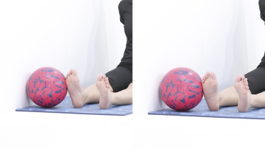 Isometric ankle eversion
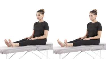 Active ankle eversion
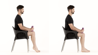 Seated calf raise
Stage 2: Strengthen the muscles that turn the foot out
When everyday walking is comfortable and stage 1 no longer brings on pain. Ankle eversion with a band makes the peroneal muscles work through a movement against resistance: turn the foot out steadily and let it come back slowly, without the band pulling it in. The band exercise for pointing the foot down and calf raises on both feet build the push-off strength you need for walking and running. Tandem stance starts the balance work, next to a counter or sturdy chair you can hold.
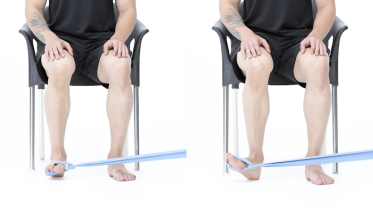 Ankle eversion with band
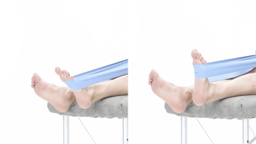 Ankle plantar flexion with band
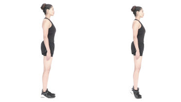 Calf raises
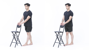 Tandem stance
Stage 3: Single leg strength and balance
When 2 to 3 sets of the band exercise and of calf raises on both feet feel easy, and the outside of your ankle is no worse the next morning. In its advice on rehab after surgery, the European expert consensus lists a single leg heel raise among the things to check before running (van Dijk and colleagues, 2018), so it makes a good goal for this stage too. The balance exercises train the quick ankle corrections you need on uneven ground: first on one leg with your eyes open, then with them closed, then on a cushion. Keep a counter or sturdy chair within reach for all of them.
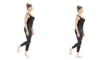 Single leg heel raise
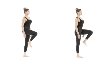 Single leg stance
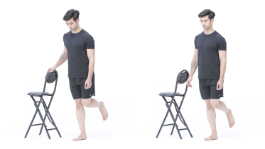 Single leg balance with eyes closed
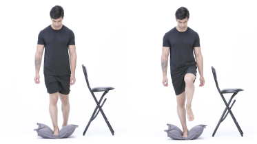 Single leg balance on a cushion
Stage 4: Hopping, then running again
When you can do about as many single leg heel raises on the sore side as on the other, balance on the cushion feels solid, and the tendon is steady the morning after your sessions. Start with low pogo hops on both feet, then hops over a line on one leg. Skater hops add side-to-side landings, which are closer to the turns in sport, so they come last. Many physios then restart running with short, easy runs on flat ground. How the tendon feels the next morning sets the pace from there. If you have osteoporosis or have had a fall in the past year, ask your physio before you do any hopping. If you have had a hip or knee replacement, get the go-ahead from your surgeon before you try hopping. Many surgeons advise against jumping and hopping after a joint replacement for good, not just for the first months, because each landing sends high impact through the new joint. If you are pregnant, have had a baby in the past year, or leak urine when you jump, cough or sneeze, check with your midwife or physio before you start this stage. With a heart condition or high blood pressure, check with your doctor before you start.
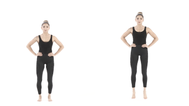 Pogo hops
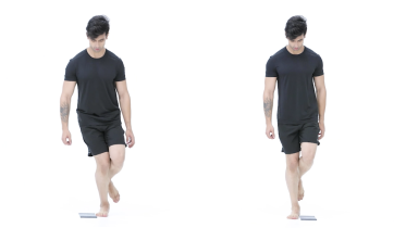 Single leg hops over a line
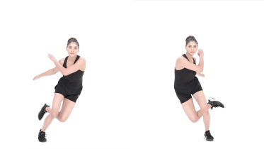 Skater hops
Getting back to running
There is no tested return to running plan for peroneal tendinopathy. The European consensus, writing about rehab after surgery on these tendons, advises that running should not start at a set time. Instead, it depends on whether you can do a single leg heel raise, and on your strength and how well you control and balance on that leg (van Dijk and colleagues, 2018). Physios often use the same idea without surgery.
Many people start with short, easy runs on flat, even ground once they can do single leg heel raises and the hopping and balance work feels solid. Build the distance before the speed. Leave hills and trails until last, along with sharp turns. Keep the strength and balance work going 2 to 3 times a week after you are back. If the tendon is clearly worse the next morning, drop back to your last comfortable level.
Snapping tendons and tears
Two related problems need a different plan. In peroneal subluxation or dislocation, the band that holds the tendons in their groove has been stretched or torn, and the tendons slide out over the ankle bone, often with a snap. The EFORT review describes the usual cause as a forceful bend of the ankle upward while the heel turns in and the peroneal muscles are working (Davda and colleagues, 2017). Exercise does not fix this on its own.
For a new dislocation, the European consensus says people who are not athletes may be offered a cast or boot for about 6 weeks, but should be told there is about a 50% chance it happens again, and it recommends surgery for elite athletes (van Dijk and colleagues, 2018). The tendons can also split or tear, most often the peroneus brevis. The consensus advises treating a tear only if it causes symptoms, with care without surgery first and surgery if that fails. A doctor or physio will usually use an ultrasound or MRI scan to tell these problems apart.
When to see a physio or doctor
See a physio or doctor if pain behind the outer ankle bone is not settling after a few weeks of easing the load, if it stops you walking normally, or if you are not sure it is coming from the tendons. A physical therapist (physiotherapist) can examine the tendons and ligaments and look at your foot shape. They can then pick your starting stage and adjust the load as you go. If the tendons snap out of place, or the pain has not improved after several months of steady treatment, ask to see a foot and ankle specialist.
After a new ankle injury, get it checked for a fracture first if you could not walk on it. Each warning sign below says how quickly to get help.
For physiotherapists
This page gives patients a starting framework for peroneal tendinopathy without instability of the tendons. Examination in the EFORT review includes foot type (cavus or planovalgus), palpation of the tendons in the retromalleolar groove during resisted dorsiflexion and eversion, and testing of the lateral ligaments, with pain on resisted eversion, passive inversion stretch or resisted plantarflexion of the first ray (Davda and colleagues, 2017). The review also lists anatomical variants such as a low-lying peroneus brevis muscle belly and a peroneus quartus. The ESSKA-AFAS consensus considers ultrasound and MRI both appropriate, reserves tendoscopy for strong clinical suspicion with negative imaging, and advises the Ottawa rules for acute injuries (van Dijk and colleagues, 2018).
Consensus statements support conservative management for all patients (4.1), shockwave when initial measures fail (4.3), and do not support platelet-rich plasma (4.4). The EFORT review advises against corticosteroid injection because of the risk of rupture, and uses a short leg cast or walker boot for 6 weeks in refractory cases; the consensus rationale, by contrast, advises avoiding immobilization in acute conservative care and starting weight bearing, range of motion and tendon loading once pain allows. Chronic loading in cavovarus malalignment is named as a predisposing factor (statement 1.3), so assess hindfoot alignment and consider a lateral forefoot post.
No trial has tested a loading protocol for peroneal tendinopathy. The progression here borrows from tendinopathy loading principles and from the balance training recommendations of the 2021 JOSPT lateral ankle sprain guideline (Martin and colleagues, 2021). Base progression on the 24-hour response. For acute dislocation in non-athletes, the consensus describes 6 weeks in a cast in slight plantarflexion or a boot with a 2 cm heel wedge, then peroneal strengthening and proprioception, with a recurrence risk of about 50% (statement 7.2), and recommends surgery for elite athletes (7.3). Criteria for running after surgery are goal based: single heel rise and strength, plus neuromuscular control and proprioception.
See a doctor promptly if
- Emergency: the ankle or foot looks deformed or out of place, you heard a crack when you were injured, or the foot is numb, tingling, cold, pale or blue. Go to an emergency department straight away.
- You felt a sudden pop, snap or tearing feeling on the outside of your ankle, and since then the tendons feel as if they slip out of place behind the bony bump, the outside of the ankle is swollen and bruised, or turning your foot outward has become weak. Stop the exercises, take the weight off that foot as much as you can and get it checked the same day at an urgent care center or emergency department, even if you can still walk. The band that holds the tendons in their groove may have torn so they slip out (peroneal dislocation), or a tendon may have torn.
- You could not take 4 steps right after the injury and still cannot, or the back edge or tip of either ankle bone is tender to press. Get it checked for a fracture the same day. These are part of the Ottawa ankle rules, explained on the ankle sprain page.
- The pain is in the middle of your foot, and the bony bump halfway along the outer edge of the foot or the bony bump on the inner side of the midfoot is tender to press. Get it checked for a fracture the same day.
- You felt a pop or snap at the back of your heel or lower calf, or it felt as if someone kicked you there, even if you can still walk and push off. Go to an urgent care center or emergency department the same day, as this can be a torn Achilles tendon rather than a sprain.
- A calf or thigh that is newly swollen, warm, tender, red or darker than usual, has swollen veins that are sore to touch, or has a throbbing or cramping pain that does not fit your injury or condition, or that feels different from normal muscle soreness after exercise. This can be a sign of a blood clot (deep vein thrombosis), especially after surgery, an injury, time in a cast or boot, a long trip or a spell of being much less mobile than usual. Get medical advice the same day. If you are also short of breath or have chest pain, call emergency services.
- The foot is hot, red and swollen, or you have a fever or feel unwell. Get medical help the same day, and go to an emergency department if you feel very unwell. If you have a very high or very low temperature, shivering you cannot control, you are breathing very fast or finding it hard to breathe, you are confused or your speech is slurred, your skin, lips or tongue look blue, gray, pale or blotchy (on brown or black skin this can be easier to see on the palms of the hands or soles of the feet), or you have a rash that does not fade when you press it, call emergency services, as this can be sepsis.
- You have diabetes or numb feet, and one foot becomes hot, swollen or red, or changes color or shape, with or without pain. Take your weight off that foot straight away and get advice the same day from your doctor or your diabetes foot team. This can be Charcot foot, where the bones and joints of the foot are damaged and the foot can collapse. Do not exercise that foot until it has been checked. If you also have a fever or feel very unwell, or redness is spreading up your foot or leg, go to an emergency department.
- You have diabetes or numb feet and find a new blister, cut or sore on your foot, especially one you did not feel, a wound that smells or is leaking, or new pain in your foot that you cannot explain. Get it seen by your doctor or foot team the same day. If you also have a fever or feel very unwell, redness is spreading up your foot or leg, or part of your foot or a toe turns black, go to an emergency department.
- A leg or foot that is getting weaker, for example your foot drags, catches on the ground or slaps down when you walk (foot drop). Get medical advice the same day. If the weakness is getting worse by the hour, go to an emergency department.
- Your tendon pain or swelling started while you were taking a fluoroquinolone antibiotic, such as ciprofloxacin or levofloxacin, or in the months after a course. Contact your doctor straight away. The UK medicines regulator (MHRA) advises stopping the antibiotic at the first sign of tendon pain or swelling and talking to your doctor. Hold off the exercises on this page until you have been checked.
- The tendons snap, click or slide over the bony bump on the outside of your ankle when you move it, and this is new, painful or keeps happening, or the ankle feels weak or gives way. This is not an emergency, but stop any exercise that brings it on and book an assessment with a doctor or physio in the next week or two, as the tendons may be slipping out of their groove (peroneal subluxation).
- Your ankle pain comes with painful, swollen joints elsewhere, or with back pain and stiffness that is worse in the morning and at night, or both ankles became painful without an obvious reason. The same applies if you also have psoriasis, Crohn's disease or ulcerative colitis, or have had a red, painful, inflamed eye (iritis). This is not an emergency, but see your doctor in the next week or two, as swollen tendon sheaths can be a sign of an inflammatory arthritis such as rheumatoid or psoriatic arthritis.
- You have numbness, tingling, burning or loss of feeling in the foot, often along the outer edge. Book an assessment with a doctor or physio in the next week or two.
- The pain is there at night or at rest as well as with activity and keeps getting worse. See your doctor within a few days. If you have had cancer, now or in the past, or are losing weight without trying, see your doctor sooner, within a day or two, and mention it. This applies even if the pain is not getting worse. If you are being treated for cancer now, contact your cancer team the same day.
Common questions
How long does peroneal tendinopathy take to heal?
There are no good studies that give a typical recovery time for this tendon, so be wary of any exact figure. Like other tendon problems, it usually settles over weeks to months rather than days, and it can flare if you go back to full running too soon. The European expert consensus suggests shockwave therapy may be considered when the first measures fail, and notes it has been suggested for tendon pain in the leg that lasts beyond 3 months (van Dijk and colleagues, 2018). Your physio will adjust your program as the tendon settles.
Can I keep running with peroneal tendinopathy?
Sometimes, if you cut it back. Many physios allow some running when it causes only mild discomfort that settles soon after and the outside of the ankle is no worse the next morning. Flat, even ground is usually easier than slopes, trails or running on a camber, and turning and cutting sports are harder than straight running. If running makes the tendon clearly worse the next day, stop it for now and keep going with the strength work.
Is peroneal tendinopathy the same as an ankle sprain?
No, although one often follows the other. A sprain is a sudden injury to the ligaments in front of and below the outer ankle bone. Peroneal tendinopathy is pain in the tendons behind that bone and along the outer side of the heel, and it usually builds up with activity over weeks. The European consensus describes peroneal tendon problems as a serious cause of pain at the back and outside of the ankle after sprains (van Dijk and colleagues, 2018), so it is worth checking the tendons if a sprain is not settling as expected.
Why does the outside of my ankle click or snap?
The two peroneal tendons are held in a groove behind the outer ankle bone by a band of tissue. If that band has been stretched or torn, often by a forceful twist of the ankle, the tendons can slide out of the groove and snap back (Davda and colleagues, 2017). If the tendons snap, click or slide over the bony bump on the outside of your ankle when you move it, and this is new, painful or keeps happening, or the ankle feels weak or gives way, stop any exercise that brings it on and book an assessment with a doctor or physio in the next week or two, as the tendons may be slipping out of their groove (peroneal subluxation). That part is not an emergency. One pattern is faster: if you felt a sudden pop, snap or tearing feeling on the outside of your ankle, and since then the tendons feel as if they slip out of place behind the bony bump, the outside of the ankle is swollen and bruised, or turning your foot outward has become weak, stop the exercises, take the weight off that foot as much as you can and get it checked the same day at an urgent care center or emergency department, even if you can still walk. The band that holds the tendons in their groove may have torn so they slip out (peroneal dislocation), or a tendon may have torn. Exercise alone does not hold the tendons back in the groove.
Do insoles help peroneal tendinopathy?
They may, especially if you have high arches or a heel that tips inward. The EFORT review lists an insole with a wedge under the outer edge of the forefoot among the first treatments in mild cases, alongside rest, activity changes and anti-inflammatory medicine (Davda and colleagues, 2017). That advice comes from expert opinion rather than trials. A physio or podiatrist can check your foot shape and advise.
Are steroid injections used for peroneal tendinopathy?
They are not a first choice. The EFORT review warns that a steroid injection carries a risk of the tendon tearing, and the authors do not use it in their own hospital (Davda and colleagues, 2017). The European consensus found no support in the research for platelet-rich plasma injections either (van Dijk and colleagues, 2018). Any injection is a decision to make with your doctor.
References
- van Dijk PA, Miller D, Calder J, DiGiovanni CW, Kennedy JG, Kerkhoffs GM, Kynsburtg A, Havercamp D, Guillo S, Oliva XM, Pearce CJ, Pereira H, Spennacchio P, Stephen JM, van Dijk CN. The ESSKA-AFAS international consensus statement on peroneal tendon pathologies. Knee Surgery, Sports Traumatology, Arthroscopy. 2018;26(10):3096-3107. https://doi.org/10.1007/s00167-018-4971-x
- Davda K, Malhotra K, O'Donnell P, Singh D, Cullen N. Peroneal tendon disorders. EFORT Open Reviews. 2017;2(6):281-292. https://doi.org/10.1302/2058-5241.2.160047
- Martin RL, Davenport TE, Fraser JJ, et al. Ankle Stability and Movement Coordination Impairments: Lateral Ankle Ligament Sprains Revision 2021. Journal of Orthopaedic and Sports Physical Therapy. 2021;51(4):CPG1-CPG80. https://doi.org/10.2519/jospt.2021.0302
- Medicines and Healthcare products Regulatory Agency. Fluoroquinolone antibiotics: must now only be prescribed when other commonly recommended antibiotics are inappropriate. Drug Safety Update. 22 January 2024. https://www.gov.uk/drug-safety-update/fluoroquinolone-antibiotics-must-now-only-be-prescribed-when-other-commonly-recommended-antibiotics-are-inappropriate
- NHS. Deep vein thrombosis (DVT). Page last reviewed 30 April 2026. https://www.nhs.uk/conditions/deep-vein-thrombosis-dvt/
Written and checked by the PocketPhysio editorial team. Last updated 2026-10-02.
This page is general education, not a diagnosis or a personal treatment plan. See a physiotherapist or doctor for advice about your own situation.