
LCL injury exercises and recovery
Warning signs, and when to get help
What is an LCL injury?
The lateral collateral ligament is a narrow, cord-like band on the outer side of your knee. OrthoInfo describes it running from the thigh bone to the fibula, the slimmer of the two bones in your lower leg, and together with the ligament on the inner side it controls the side-to-side movement of the joint. Its job is to stop the knee opening outward.
The usual cause is a force on the inner side of the knee that pushes it outward. ChoosePT adds two more: the knee being straightened too forcefully, and a quick change of direction or a sharp turn with the foot planted. Most of these happen in sport, and that guide says LCL sprains affect women and men equally. Its age figures cover collateral ligament knee injuries as a group, not this ligament alone, and they fall into two bands: adults aged 20 to 34, and adults aged 55 to 65.
What people notice afterward is pain, swelling and tenderness along the outer side of the joint, often with a pop at the moment it happened, and a knee that feels as though it could give way. OrthoInfo lists instability and stiffness as well, including not being able to fully straighten or bend the knee. If the pain sits along the outer side without an injury behind it and built up with running, IT band syndrome and the other causes on outer knee pain fit better than this page.
Doctors and physios grade the sprain by how far the knee opens when they push it. OrthoInfo describes grade 1 as mildly damaged and slightly stretched but still able to help keep the joint stable, grade 2 as stretched to the point where the ligament becomes loose, which it calls a partial tear, and grade 3 as torn in half or pulled directly off the bone, with the joint unstable.
This page is for adults with an injury to this ligament managed without an operation, including people still deciding. If a surgeon has seen your scans, confirmed that the tear is complete but involves this ligament alone, and decided against operating, then the plan that came with that decision is the one to follow, not this one. The week off weight bearing and the hinged brace OrthoInfo describes are written for a stable grade 1 or grade 2 injury, a complete tear is protected for longer than that, and how much longer is a question for the clinician who graded it. If you came out of that appointment without a plan, which happens often enough to be worth saying, ring back and ask for one before you start anything: how long the brace stays on and what range it is set to, how much weight goes through the leg and for how many weeks, and who is going to watch the knee while it heals. Until you have those answers, keep to what you were told about the brace and the crutches and do not start the stages below.
A child or teenager who is still growing and injures a knee should be checked before starting any program. If your knee has been operated on, your surgeon's and physio's plan comes first, including where it differs from anything here.
Is it just the LCL, or the posterolateral corner too?
An injury to this ligament on its own is uncommon, and that one fact changes how your knee should be assessed. OrthoInfo says these account for less than 2% of knee injuries, and a review of isolated fibular collateral ligament injuries in athletes calls them rare and makes the point that checking for other injuries alongside them is the critical part of treatment (Sikka and colleagues, 2015). So the question is bigger than how badly the ligament is torn. It is what else went with it.
What often goes with it is the posterolateral corner: the group of structures at the back and outer side of the knee that work with this ligament to stop the joint opening outward and rotating. An international expert panel reached consensus on several points that matter to you as a patient. Repairing a complete tear on its own, without augmentation or reconstruction, should not be done, and a complete grade 3 corner injury should have a reconstruction, although an acute avulsion of a single structure can be repaired. Treatment has to be matched to the individual knee, with staged rehabilitation. A meniscal or nerve injury alongside the corner affects when surgery happens, and no classification system yet exists that usefully tells a surgeon what to do (LaPrade and colleagues, 2025).
That panel's statements are expert opinion, which is the level they give themselves, so read them as the shape of the decision, not as a protocol.
The nerve is the part to take seriously, and it is the reason this page spends as long on the diagnosis as on the exercises. The common peroneal nerve wraps round the top of the fibula, right where this ligament attaches, so a force that pushes the knee outward stretches the nerve along with the ligament. Between 16% and 40% of people with a multi-ligament knee injury have an injury to that nerve. Sitting that close to the bone is what leaves it exposed, and it is highly vulnerable to a stretch injury under outward force, especially in corner injuries. Separately, and this is the part worth knowing, the outlook for this nerve is poorer than for other nerve injuries in the arms and legs (Mook and colleagues, 2013).
In a series of 47 people operated on for a corner injury, 11 had a nerve palsy, and 4 of those 11 recovered fully; the palsy went with injuries at the lower end of the corner and with the hamstring tendon being pulled off the fibula (Essilfie and colleagues, 2022). Those were operated knees, not mild sprains, so do not read your own odds from them.
Behind all of that sits the worst version, and it is the time-critical one. Enough force to tear several ligaments can dislocate the whole knee, and the artery and nerve behind the joint can be damaged in the same moment. The knee can go back into place by itself and look almost ordinary afterward, with a warm, pink foot, and the person describes it as having gone out and come back. That is a hospital assessment on the day, not a physiotherapy appointment, which is why the first warning signs on this page send you to an emergency department.
That route to hospital comes from the systematic review of vascular and nerve injury after knee dislocation (Medina and colleagues, 2014). The PCL injury program and MCL sprain program carry the same warnings for the same reason.
So the fork, in plain terms. A stable grade 1 or grade 2 injury to the ligament alone follows the program below, in whatever brace you were given. A complete tear, a knee that is loose in more than one direction, or a knee that shifted out of place belongs with a knee surgeon first. And two of those signs are timed rather than merely urgent: a knee that felt as though the whole joint went out, even for a second, and any numbness, coldness or new weakness in the foot both need an emergency department on the day, not an appointment later in the week.
Which side of the fork you are on is not a thing you can read off your own knee. OrthoInfo's answer to that question is an examination of every structure in the joint, usually with imaging to confirm it and to rule out the injuries that travel with this one. If none of the signs above fits you, that is reassuring rather than proof.
What varus load is, and where you meet it
You will be told to avoid varus stress and you may well not be told what it is. Varus load means any force that pushes your knee outward, or pushes your lower leg inward underneath it, so the outer side of the joint opens and this ligament is the thing holding it shut. That is the exact direction that injured it. The following examples are reasoning from how the ligament works rather than findings from a trial, so treat them as practical rather than proven.
The obvious ones happen in sport: a tackle or a fall where someone lands against the inside of your knee, a sidestep where the knee is driven outward, and a landing on one leg where the knee travels out over the foot. Those are the moments physios ask about, and they are why contact and cutting come back last.
The quieter ones happen all day, and they are the reason a knee that was settling stops settling. Letting the knee drift outside the line of your foot as you squat, step down or stand up from a low chair, which is the same position as the landing above, only slower. Sitting cross-legged on the floor with the knee pulled out and down, which combines the outward load with a twist. Ground that tips the leg sideways without warning: a rutted field, a curb taken at an angle, a pothole, a loose stone.
On uneven ground you do not get to choose which way the knee is pushed, which is the whole problem with it. Flat, even ground and shoes that hold the foot level take most of this away, which is a dull piece of advice that changes more than any single exercise.
How long does an LCL injury take to heal?
Nobody has published a timetable for this ligament, and the strongest patient source says as much: ChoosePT states that the healing time varies with each person. What exists is the non-operative shape OrthoInfo describes for a stable grade 1 or grade 2 injury, which is about a week without weight through the leg and then a hinged brace while it heals, with physical therapy alongside.
So use the order rather than the calendar. The limp goes first, then stairs stop needing the handrail, then you stop thinking about the knee in a crowd. Strength and confidence on one leg come after that, and sideways movement comes last, which is why this program ends where it does. A private marker worth watching for is the moment you step down a curb without planning it.
Two honest warnings about numbers. The brace coming off is not the finish line, because the ligament healing and the leg being strong are weeks apart, and the gap is the whole of stages 3 and 4. And a complete tear, or one that turns out to be part of a corner injury, runs on a different clock set by a surgeon rather than by this page.
How to use this program
Start at stage 1 if the injury is recent or the knee is still swollen, and at stage 2 or 3 if this happened a while ago and the knee is only unreliable under load. If you are not sure, start at stage 1. Move up when the stage you are on feels easy and the knee is no worse the morning after. Those move-up rules are common practice rather than tested rules, so use them as a guide. The gates a few paragraphs below are a different thing: those are conditions, not a guide.
Each exercise page gives its own starting dose, and the stage texts above repeat the numbers so you can plan a session without opening four pages. Across the program that is mostly 2 to 3 sets of 8 to 15, with the still holds at 5 to 10 seconds, the balance holds at 10 to 30 seconds and the foam pad at 20 to 30 seconds. The lying and sitting work can be done daily and the harder standing work 2 or 3 days a week so the muscles recover. Some of the exercise pages offer the standing and balance work once or twice a day, because they are written for knees in general: on this knee use the 2 or 3 days a week above and not the daily option, and do not add the two together. Your physio will adjust the exercises and the numbers to your knee and your sport.
Before you start, read these gates and check them against yourself. They sit here, above the program, because a reader who scrolls to the exercises and starts is the one they are written for.
Has the knee been examined for other ligament damage? An isolated sprain and a corner or multi-ligament injury are not the same problem and they do not share a plan. If nobody has pushed and pulled your knee in several directions, that comes before stage 1.
Were you given a brace or crutches? If so, this program runs inside your clinician's instructions for them, not around them. Wear the brace as you were told to and follow their limits on how much weight goes through the leg, even where that means doing less than a stage says.
If you have had any knee surgery, a repair, a reconstruction, a cartilage trim or a replacement, your surgical team's own instructions replace this program. Anything new in an operated knee goes to that team the same day rather than to a web page: new pain, swelling, heat or giving way all need ruling out before anyone treats them as a training error.
If you have fallen in the past year, feel unsteady on your feet, or take medicines that make you dizzy, keep a hand on a kitchen counter through the standing and balance work, practice only when someone else is at home, and ask your doctor or physio about a falls check. If you live alone, plan around that rather than ignoring it: keep to the lying and seated exercises when you are on your own, do the standing work at your appointments or in a class, and ask your physio which of them you may do alone.
If you have high blood pressure, breathe steadily through every repetition and never hold your breath. Check with your doctor before you start if you have a heart or lung condition, or blood pressure that is not under control. If you are pregnant, the exercises done lying on your front or flat on your back need changing as pregnancy goes on, so plan those with your physio and your maternity team.
Four of the warning signs below stop this program outright, and all four send you to an emergency department on the day rather than to an appointment, which is the reason to read that list now rather than later: a foot that has dragged, caught, slapped or been numb on top since the injury itself; a knee that felt as though the whole joint shifted out of place, even if it went straight back; a knee loose or wobbly in more than one direction; and a whole knee that swelled up within a few hours of a twist, a fall or a blow, or a pop or a snap at the moment it happened. A foot that has slowly got weaker since, rather than from the moment it happened, is a same-day call instead, and the list says so.
The exercise program
The exercises below carry their own stop signs and precautions. Open the line under an exercise and read them before you try it.
Stage 1: Protect the ligament and switch the thigh back on
The first days and weeks, while the outer knee is sore and you may be in a brace or on crutches. Wear the brace you were given and follow your clinician's instructions for how much weight goes through the leg, because those come before anything on this page. Everything here is done lying or sitting, so nothing pushes the knee sideways. Quad sets and short arc quads work the last part of straightening, which is the part a swollen knee gives up first, hamstring sets give the back of the thigh something to do before resisted bending is comfortable, and the heel slides get the bend moving again. The exercise pages put the quad sets and hamstring sets at about 10 holds of 5 to 10 seconds several times a day, short arc quads at 2 to 3 sets of 10 to 15 with a 3 to 5 second hold once or twice a day, and the heel slides at 10 to 15 slow repetitions 2 or 3 times a day. Your physio will adjust this, and after an operation your surgeon's and physio's plan replaces these numbers. One of these four needs naming for an operated knee. If your knee was reconstructed, leave the hamstring set out until your surgeon or physio tells you it is time, because digging the heel down works the hamstring on its own, and after a reconstruction of the posterolateral corner that kind of work is held back for a minimum of four months. Slide the heel only as far as it goes comfortably, and keep this stage pain free.
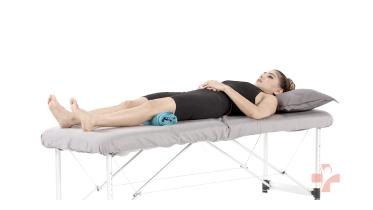 Quad sets (towel under knee)
Stop signs and precautions for the quad sets (towel under knee)
Before you start
- After a knee fracture or knee surgery, including a knee replacement, follow your surgeon's or physio's plan, and start this exercise only when they have cleared you.
When to stop and get help
- Your knee pain goes up sharply.
- Your calf or thigh is getting more swollen, or becomes warm, tender, red or darker than usual, or has a new throbbing or cramping pain. This can be a blood clot, especially after an operation, an injury, time in a cast, boot or brace, or a spell of being much less mobile than usual, and after an operation it can also be an infection. Stop and contact your medical team or get medical advice the same day. If you are also short of breath or have chest pain, call emergency services.
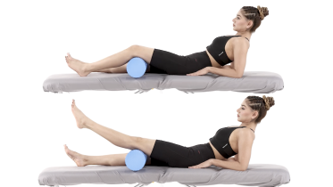 Short arc quads
Stop signs and precautions for the short arc quads
Before you start
- After a knee fracture or knee surgery, including a knee replacement, follow your surgeon's or physio's plan, and start this exercise only when they have cleared you.
When to stop and get help
- Knee pain increases sharply during the set.
- Your calf or thigh is getting more swollen, or becomes warm, tender, red or darker than usual, or has a new throbbing or cramping pain. This can be a blood clot, especially after an operation, an injury, time in a cast, boot or brace, or a spell of being much less mobile than usual, and after an operation it can also be an infection. Stop and contact your medical team or get medical advice the same day. If you are also short of breath or have chest pain, call emergency services.
- Your wound becomes more red, hot, swollen or painful, or starts to leak. Contact your medical team the same day.
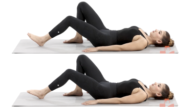 Hamstring sets (heel dig)
Stop signs and precautions for the hamstring sets (heel dig)
Before you start
- With a recent hamstring strain, even a still hold loads the injured muscle, so let your physio set when you start and how hard you press. The same applies after an ACL reconstruction that used a hamstring graft, where hamstring work is held back while the place the tendon was taken from heals, and the date comes from your surgeon's protocol. If you are pregnant and beyond the first trimester, ask your physio before you do this lying on your back.
- After a reconstruction of the posterolateral corner, the structures at the back of the outer side of the knee, leave this one out until your surgeon or physio tells you it is time. Digging the heel down to squeeze the back of the thigh is hamstring strengthening on its own, even though nothing moves and nothing resists you, and the protocol most widely followed holds hamstring strengthening on its own back for a minimum of four months, because the hamstring tendon on the outer side pulls on the fibular head, the small bone the graft is anchored into. So expect months rather than weeks there. If you have not had knee surgery, that wait does not apply to you.
- After an injury to the ligament at the back of the knee (the posterior cruciate ligament), the hamstrings pull the shin backward, which is the direction this ligament holds, so hamstring work is usually kept out of the early weeks. The knee stays close to straight in this hold, which is not where that pull is strongest, but your physio decides whether and when it fits, so do not start it on your own.
When to stop and get help
- Cramp in the back of the thigh, or a sudden sharp pull there. Stretch the leg out along the floor and rest.
- Knee pain that climbs sharply during or after the holds.
- Your calf or thigh is getting more swollen, or becomes warm, tender, red or darker than usual, or has a new throbbing or cramping pain. This can be a blood clot, especially after an operation, an injury, time in a cast, boot or brace, or a spell of being much less mobile than usual, and after an operation it can also be an infection. Stop and contact your medical team or get medical advice the same day. If you are also short of breath or have chest pain, call emergency services.
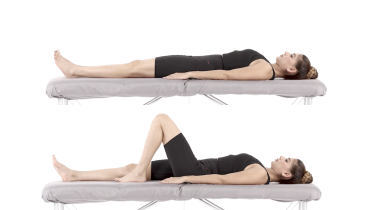 Heel slides
Stop signs and precautions for the heel slides
Before you start
- After a knee fracture or knee surgery, including a knee replacement, follow your surgeon's or physio's plan, and start this exercise only when they have cleared you.
- If you have had a hip replacement or other hip surgery, get the go-ahead from your surgeon or physio before you try heel slides. Sliding the heel toward you bends the hip, and many surgeons set a limit on how far it can bend for the first months after some operations, so find out what yours is.
- If you are pregnant and past the first three months, check with your physio before exercising flat on your back.
When to stop and get help
- Pain in the knee or hip becomes sharp or keeps increasing with each rep.
- Your calf or thigh is getting more swollen, or becomes warm, tender, red or darker than usual, or has a new throbbing or cramping pain. This can be a blood clot, especially after an operation, an injury, time in a cast, boot or brace, or a spell of being much less mobile than usual, and after an operation it can also be an infection. Stop and contact your medical team or get medical advice the same day. If you are also short of breath or have chest pain, call emergency services.
- Your wound becomes more red, hot, swollen or painful, or starts to leak. Contact your medical team the same day.
Stage 2: Strength with weight on the leg
Move up when you walk without crutches and without a limp, with or without the brace, you can lift the leg straight without the knee sagging, and stage 1 feels easy. Two things to settle before you start this stage, rather than after, because both of them can change what you do in it. Ask your physio before you add the band to the hamstring curl if nobody has examined this knee yet, or if you were told more than this one ligament was injured: in the more severe corner injuries one of the hamstring tendons is pulled off the small bone on the outer side of the knee, and resisted bending pulls on that tendon. If the injury is a sprain of this ligament alone, resisted bending is not a sideways load on it and the band is a normal part of the stage. And if you still wear a brace, or you have had an operation on this knee, ask before you start the band and the wall squat at all, and leave both out of your sessions until you have that answer rather than starting them while you wait. After a reconstruction of the posterolateral corner, the group of structures at the back of the outer side of the knee, hamstring work on its own is commonly kept out of the program for at least four months, because the hamstring tendon pulls on the small bone the graft is anchored into, so expect that answer to be months rather than weeks away. If your operation reconstructed this ligament by itself, the graft sits in that same small bone, but nobody has published a timetable for that operation on its own, so the wait is whatever your surgeon says and this page will not put a number on it. The glute bridge builds the buttock without putting the knee under load. The mini squat and the wall squat then load the leg with your foot planted, which is the friendlier direction for this ligament, because the thigh muscles and the joint surfaces share the work and nothing is pulling the knee sideways. The lying hamstring curl with a band adds resistance to bending the knee. Keep the kneecap tracking over your second toe with the whole foot flat, and do not let the knee drift outside the line of your foot, which is the position that opens the outer side of the joint. Common numbers are 2 to 3 sets of 10 to 12 bridges with a short hold, 2 to 3 sets of 10 to 15 mini squats once a day, 2 to 3 sets of 8 to 12 slow wall squats a few days a week at a depth you control, and 2 to 3 sets of 10 to 15 curls on each leg with a light band. Your physio will adjust this.
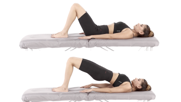 Glute bridge
Stop signs and precautions for the glute bridge
Before you start
- After hip, knee or back surgery, your surgeon's protocol decides when you can start bridges. Once you are past the first three months of pregnancy, check with your physio before exercising flat on your back.
When to stop and get help
- You feel back pain rather than buttock work, even when you lift a little lower.
- Pain spreads down the leg or you get numbness or tingling.
- Your hamstrings (the back of the thighs) cramp. This is a common complaint with bridges, and bringing your feet a little closer to your bottom often eases it.
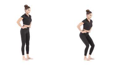 Mini squat
Stop signs and precautions for the mini squat
Before you start
- If you have had a hip replacement or other hip surgery, get the go-ahead from your surgeon or physio before you try mini squats. Some operations limit this kind of work for the first months, so the timing depends on your surgery.
When to stop and get help
- Knee pain is sharp or keeps getting worse through the set.
- The knee swells, gives way or feels unstable afterwards.
- You feel dizzy, lightheaded, faint or sick, or the room seems to spin. Hold your support, sit down and get medical advice the same day before you practice again. If you faint, it does not settle within a few minutes of sitting still, your vision suddenly blurs or goes double, or you cannot stand or walk steadily, call emergency services.
- You feel unsteady enough that you might fall. Hold on to something firm and sit down.
- Your calf or thigh is getting more swollen, or becomes warm, tender, red or darker than usual, or has a new throbbing or cramping pain. This can be a blood clot, especially after an operation, an injury, time in a cast, boot or brace, or a spell of being much less mobile than usual, and after an operation it can also be an infection. Stop and contact your medical team or get medical advice the same day. If you are also short of breath or have chest pain, call emergency services.
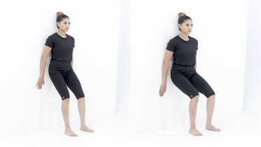 Wall squat
Stop signs and precautions for the wall squat
Before you start
- If you have had a hip or knee replacement, get the go-ahead from your surgeon or physio before you try wall squats. Sliding down until your thighs are level bends the hip to a right angle or more, and many surgeons ask you to avoid that for the first months after some operations. Ask them how deep you may go.
- If you are pregnant, check with your midwife or physio before you start.
- If you have high blood pressure, keep the holds short and breathe out as you push back up.
When to stop and get help
- Sharp pain in or around the kneecap, or pain that keeps building through the set.
- The knee gives way, locks, or swells after the session.
- Lower back or hip pain that gets worse as you slide down.
- You are pregnant and notice bleeding or fluid leaking from your vagina, regular painful tightenings, tummy pain that does not settle when you rest or comes with lower back pain, or a change in your baby's usual movements, such as moving less than usual. Stop and contact your midwife or maternity unit straight away. Do the same for a severe headache, problems with your vision such as blurring or flashing lights, pain just below your ribs, or sudden swelling of your face, hands or feet, which can be signs of pre-eclampsia. Call emergency services if the bleeding is heavy, the tummy pain is severe, bleeding or tummy pain comes with pain in your shoulder or feeling sick, faint or dizzy, or a headache comes on suddenly and is very severe.
- You feel dizzy, lightheaded, faint or sick, or the room seems to spin. Stop, sit down and get medical advice the same day before you exercise again. If you faint, it does not settle within a few minutes of sitting still, your vision suddenly blurs or goes double, or you cannot stand or walk steadily, call emergency services.
- Chest pain or pressure, dizziness or feeling faint, a racing or irregular heartbeat, or being far more out of breath than the effort should cause.
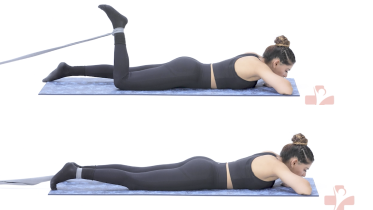 Lying hamstring curl with band
Stop signs and precautions for the lying hamstring curl with band
Before you start
- The band turns this into resisted hamstring work, which is held back after knee surgery until your surgeon or physio clears it, so ask before you use it, whatever the operation was. How long you wait depends on which operation that was. After an ACL reconstruction using a hamstring graft the wait protects the place the tendon was taken from, and the date comes from your surgeon's protocol. After a reconstruction of the posterolateral corner, the structures at the back of the outer side of the knee, the protocol most widely followed holds isolated hamstring work back for a minimum of four months, because the hamstring tendon on the outer side pulls on the fibular head, the small bone the graft is anchored into, so expect months rather than weeks there. If you have not had knee surgery, these waits do not apply to you.
- After an injury to the ligament at the back of the knee (the posterior cruciate ligament), curls like this one are usually kept out of the early weeks, because the hamstrings pull the shin backward and that is the direction this ligament holds. Ask your physio before you start it.
- If you have had recent heart or chest surgery, do not lie face down on a healing chest wound. The weight of your chest presses on the wound for as long as you are down there, and pushing up through your arms to get off the bed pulls across the breastbone. Ask your surgeon or surgical team when lying on your front is allowed, and until then ask your physio for another way to do this while you lie on your side, sit or stand. If the breastbone clicks, grinds, shifts or feels as if it is moving while you do it, stop, and contact your surgical team the same day.
When to stop and get help
- Sharp pain behind the knee or high at the back of the thigh near the buttock.
- A sudden sharp pain, pop or snap at the back of your thigh. Stop straight away. If you then get large bruising spreading down the back of the thigh, a gap or dip you can feel, a stiff-legged walk, or clear weakness when you bend the knee, get assessed the same day at urgent care or an emergency department, as the tendon may have pulled away from the bone near the buttock.
- Lower back pain while you lie or lift.
Stage 3: Balance, sideways movement and stairs
Start when stage 2 feels easy and the knee is no worse the morning after. This is the first stage that moves you sideways and the first that takes the ground away from under you, which is on purpose: an outer knee that has been stretched tends to feel unreliable on uneven ground long after it stops hurting. Keep the side steps small at first. The foam pad has both feet on it, so it is the gentle way to meet a soft surface rather than a test. Use a low step for the step up, about the height of one stair, and push through the whole foot rather than hauling yourself up with the back leg. Set every one of these up beside a kitchen counter or a rail you can reach at once, on a clear floor with no loose rugs, and wear flat shoes that grip rather than socks or bare feet. Many programs use 2 to 3 lengths of 10 to 15 side steps each way, 3 to 5 holds of 20 to 30 seconds on the pad, and 2 to 3 sets of 8 to 12 step ups on each leg. Your physio will adjust this and set the step height.
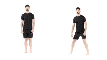 Side stepping
Stop signs and precautions for the side stepping
Before you start
- If you have had a hip replacement or other hip surgery, get the go-ahead from your surgeon or physio before you try side stepping. Some operations limit this kind of work for the first months, so the timing depends on your surgery. If the operation was done through the side of the hip (a lateral approach), the muscles at the side of the hip were detached and repaired. Exercises that make them work hard, like this one, are then often held back for around the first 6 weeks.
When to stop and get help
- You feel dizzy, lightheaded, faint or sick, or the room seems to spin. Hold your support, sit down and get medical advice the same day before you practice again. If you faint, it does not settle within a few minutes of sitting still, your vision suddenly blurs or goes double, or you cannot stand or walk steadily, call emergency services.
- You start to lose your balance.
- Sharp pain in the hip, groin or knee.
- Pain running down the leg, or the leg going numb or tingly.
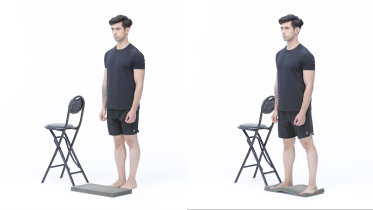 Standing balance on a foam pad
Stop signs and precautions for the standing balance on a foam pad
When to stop and get help
- You feel dizzy, lightheaded, faint or sick, or the room seems to spin. Hold your support, sit down and get medical advice the same day before you practice again. If you faint, it does not settle within a few minutes of sitting still, your vision suddenly blurs or goes double, or you cannot stand or walk steadily, call emergency services.
- You start to fall and need the support to stop yourself.
- Sharp pain in an ankle or knee, or an ankle that gives way.
- Your feet feel newly numb or tingly, or your balance has been slipping over recent weeks. Get medical advice within a few days, and leave out eyes-closed work until you have been checked. If the numbness or tingling spreads quickly up your legs or into your hands, get medical advice the same day. If your legs also feel stiff, heavy or weak, walking suddenly becomes hard, you have trouble breathing or swallowing, or you have trouble controlling your bladder or bowels, call emergency services.
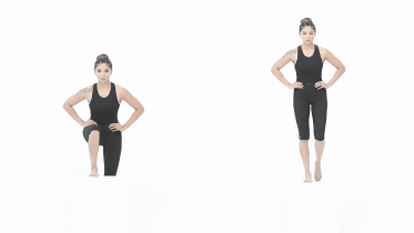 Step up
Stop signs and precautions for the step up
Before you start
- If you have had a hip or knee replacement, or other hip surgery, get the go-ahead from your surgeon or physio before you try step ups. Some operations limit this kind of work for the first months, so the timing depends on your surgery. Your surgeon or physio will also tell you what step height is right for you.
When to stop and get help
- You feel sharp pain in the knee or the knee gives way.
- Pain at the front of the knee builds with each rep.
- You feel dizzy, lightheaded, faint or sick, or the room seems to spin. Hold your support, sit down and get medical advice the same day before you practice again. If you faint, it does not settle within a few minutes of sitting still, your vision suddenly blurs or goes double, or you cannot stand or walk steadily, call emergency services.
- You feel unsteady enough that you might fall. Stop and sit down.
- Chest pain or pressure, dizziness or feeling faint, a racing or irregular heartbeat, or being far more out of breath than the effort should cause.
Stage 4: One leg, and the control sport asks for
Start when stage 3 feels easy, you can stand on the injured leg on a firm floor for about 30 seconds without holding on, and you can go up and down a step on that leg without the knee wobbling or the hip on that side dropping. If you cannot do those yet, the work is still in stage 3, and this stage will only teach you to hold the rail. This is the last stage, and the one most people stop short of. Both exercises put you on one leg, which is how you actually land, turn and step down a curb. The cushion is a harder step than a firm floor because the surface keeps moving, so the ankle answers in quick small corrections and the hip tidies up the rest. In the lateral step-down the band pulls your knee inward and you hold it in line with your second toe, which trains the hip to control the knee under load. That inward pull is worth understanding on this page: it is the opposite direction to the one that injured your outer ligament, so it is a reasonable exercise here, and the useful part is the single leg control rather than the band itself. Start with a light band, a low step and a wall or rail within reach, and if the kneecap leaves the line of your second toe before you are halfway down, the band is too strong or the step is too high. Usual numbers are holds of 10 to 30 seconds 2 to 3 times on each leg, and 2 to 3 sets of 8 to 12 step-downs on each leg. Your physio will adjust this. Running, cutting, jumping and contact come after this stage, on a plan you agree with your physio, and a knee that still feels as though it could open up sideways is not ready for them.
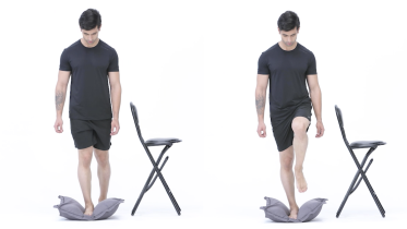 Single leg balance on a cushion
Stop signs and precautions for the single leg balance on a cushion
When to stop and get help
- You feel dizzy, lightheaded, faint or sick, or the room seems to spin. Hold your support, sit down and get medical advice the same day before you practice again. If you faint, it does not settle within a few minutes of sitting still, your vision suddenly blurs or goes double, or you cannot stand or walk steadily, call emergency services.
- You lose your balance and have to grab the chair hard to stay up.
- Your ankle gives way, or you feel sharp pain in the ankle or knee.
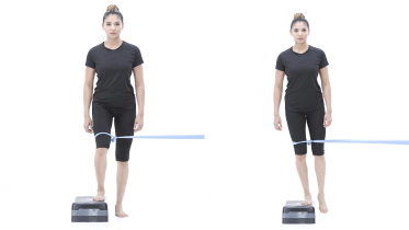 Lateral step-down with band
Stop signs and precautions for the lateral step-down with band
Before you start
- If you have had a hip replacement or other hip surgery, get the go-ahead from your surgeon or physio before you try lateral step-downs. Some operations limit this kind of work for the first months, so the timing depends on your surgery. After knee surgery, wait until your surgeon and physio clear you for single leg step work.
When to stop and get help
- Sharp pain in the knee, or the knee buckles or gives way.
- Pain at the front of the knee that grows rep after rep.
- You feel unsteady enough to fall. Hold the wall or rail and step off.
- You feel dizzy, lightheaded, faint or sick, or the room seems to spin. Hold your support, sit down and get medical advice the same day before you practice again. If you faint, it does not settle within a few minutes of sitting still, your vision suddenly blurs or goes double, or you cannot stand or walk steadily, call emergency services.
- Chest pain or pressure, dizziness or feeling faint, a racing or irregular heartbeat, or being far more out of breath than the effort should cause.
Getting the detail right
Stage 1 is quiet work that pays off later, so do it properly. In quad sets, press the back of the knee down into the towel and watch the kneecap slide a short way toward your hip, instead of squeezing the whole leg and holding your breath. If the kneecap sits still while your buttock grips, the thigh has not come on. Short arc quads want a rolled towel or roller under the knee and the heel lifting only until the knee is straight, with the back of the knee staying on the roll; if the roll shifts, the hip has taken over. In heel slides, a sock on a wooden floor or a plastic bag under the heel makes the slide honest, and little and often beats one hard attempt.
In stage 2 the outer knee is testing you sideways even when the exercise looks straight. For the mini squat and the wall squat, keep the kneecap tracking over the second toe with the whole foot flat on the floor. The knee drifting out past the line of the foot is the thing to catch, and almost nobody notices it happening, so face a mirror for a set. In the lying hamstring curl with a band, keep the hips pressed into the mat and pick a band light enough that you still reach the top of every repetition.
Stage 3 is where the honest wobble shows up. Keep the side steps small at first and keep your toes pointing forward across the room instead of turning them the way you are heading, which turns it back into ordinary walking. On the foam pad, start firmer than you think, because a deep squashy camping mat gives way so much that the only thing getting a workout is your grip on the counter. Use a low step and watch the working knee on the way up.
Stage 4 is about control rather than effort. Go slowly enough in the lateral step-down that you could stop halfway down, check the band knot before every set, and set up beside a wall or a rail every single time. On the cushion, expect it to feel busier rather than heavier, and if you are grabbing the chair again and again, go back to a firm floor for a week.
After surgery for an LCL or posterolateral corner injury
If your ligament was repaired or reconstructed, this program is not your program. Your surgical team's own protocol sets your brace, how much weight goes through the leg, how far the knee may bend and when each of those changes, and their numbers come first everywhere they differ from this page.
This page will not set those numbers for you, and the reason goes beyond caution. The expert panel above found that no single reconstruction technique is universally accepted and that treatment and staged rehabilitation have to be individualized to the knee (LaPrade and colleagues, 2025). A technique paper on posterolateral corner reconstruction makes the same point from the surgeon's side, noting that the operation is technically demanding, that injury to the common peroneal nerve is a recognized risk of it, and that in longstanding cases an unrecognized problem with the alignment of the leg can be why a reconstruction fails, so the standing alignment of the whole leg is assessed beforehand (Maheshwer and colleagues, 2022). A protocol built around one surgeon's technique does not transfer to another's, and a web page cannot know which you had.
There is still a common shape to the first few months, and it is worth knowing so that you have something to ask about rather than nothing. Read what follows knowing what it is. It is one published protocol, written by one group of surgeons around their own technique, which many other surgeons follow. It is not a standard, it is not a schedule, and it is not your plan. Your own dates will differ from it somewhere, and that is ordinary rather than a sign that something has gone wrong.
In that protocol, the knee stays in an immobilizer, or a brace set in full extension, with all weight off the leg for about six weeks. Bending is done passively and kept to about 90 degrees for the first two weeks, then opens up as the knee allows. Crutches are weaned and a stationary bike started at around six weeks, once weight is allowed through the leg. Strength work starts with the foot planted. Hamstring work on its own is held back for at least four months. Running and agility wait until strength and power are back, usually around six months, and return to sport is commonly six to nine months (Chahla and colleagues, 2016; Shon and colleagues, 2017).
Both of those reviews say the numbers get changed when other ligaments were reconstructed at the same time, which is most of these knees. So use them as the questions to put to your own team, and for nothing else. If your team's dates differ from these in either direction, theirs are the right ones for your knee: being further on than this page is not a danger sign, and being behind it is not a failure. And do not take a number here as a reason to do less than your team has asked, or to stay off the leg longer than they told you to, because the time off the leg is set for your own repair and the thigh muscle fades fast while you wait.
One of those limits lands directly on the program above, so it is worth spelling out. Stage 2 uses a lying hamstring curl against a band and stage 1 uses a hamstring set, and after a reconstruction both of those are exercises to leave alone until your surgeon tells you otherwise. The hamstring tendon on the outer side of the thigh pulls on the fibular head, the small bone on the outer side of the knee, which is exactly where the graft is anchored. That is why hamstring strengthening on its own is kept out of the corner reconstruction protocol for a minimum of four months after the operation (Chahla and colleagues, 2016; Shon and colleagues, 2017).
Bending the knee as a movement is not the problem. The muscle pulling on that bone is, and it pulls whether the resistance comes from a band, a weight or the bed under your heel.
Neither review says whether a still hold counts as hamstring work on its own, so this page will not clear the stage 1 hamstring set for you either: ask about that one as well, rather than treating it as the safe version of the same thing. Neither review sets a time for a reconstruction of this ligament by itself either, so if that was your operation the number comes from your surgeon rather than from here. Ask before you do any hamstring work on its own, and after a corner reconstruction expect the answer to be months rather than weeks.
What this page can usefully give a post-operative reader is the watch list. The warning signs below include the lines written for an operated knee: a wound that leaks or opens, redness, heat or a fever, and a knee that gives way, pops or swells after a twist. That last line says "reconstructed" because that is the commoner operation here, and it applies in the same way if your ligament was repaired. Those are same-day calls to your surgical team rather than things to train through. A new foot drop, or new numbness on the top of the foot, after the operation belongs in that group too, because injury to that nerve is a recognized risk of this surgery: tell your surgical team the same day rather than saving it for the next appointment, and if the weakness is getting worse by the hour, the warning signs below send you to an emergency department instead.
Ask your team three questions at your next appointment, because they decide how you use the stages here afterward: how long is the brace on and what range is it set to, what is my weight bearing and bending limit this month, and when may I start single leg work.
Some of this moves faster than a same-day call, and the first sign of it is pain rather than numbness. Calf or shin pain that keeps climbing, pain far worse than you were told to expect or that your painkillers do not touch, or a dressing, bandage or brace that feels tight on the leg: any one of those on its own means an emergency department straight away, and do not drive yourself. New weakness or numbness in the foot alongside any of them makes it more urgent again rather than less, and it tends to come later rather than first, so do not wait for it before you go. Bleeding or swelling under a tight dressing can press on that nerve, and pressure building up inside the leg (compartment syndrome) has to be ruled out quickly. Take the pressure off rather than waiting to get through to your team first: loosen a brace strap or a bandage that is squeezing the leg, leave the dressing stuck over the wound alone and leave the brace itself on, keep the leg at about heart level rather than propped higher than that, and leave the ice off.
Two more things if yours was a repair rather than a reconstruction. Repair on its own has failed more often than reconstruction in the published series: in one surgeon's comparison in multi-ligament knees, 4 of the 10 repairs of this ligament and the corner failed against 1 of the 18 reconstructions (Levy and colleagues, 2010), and the expert panel's position is that a complete corner tear should not be repaired on its own without augmentation or reconstruction, while an acute avulsion of a single structure can be (LaPrade and colleagues, 2025). Read those two numbers properly before they frighten you. They are 28 knees in total, all done by one surgeon, the two groups operated on one after the other in different years rather than side by side, and every knee in them had several ligaments torn rather than this one alone. Four out of ten is not a 40 percent chance for you, in either direction, because ten knees cannot be turned into anybody's odds.
What the comparison is good for is the direction it points, which is why a complete corner tear is now reconstructed rather than repaired, and the same paper reported that once the failed repairs had been reconstructed the two groups scored about the same. So a repaired knee that starts to feel loose again, or that gives way, goes to your surgeon rather than into the next session, and it is worth asking what was repaired and whether it was augmented.
The nerve is the other thing. The common peroneal nerve is found and freed as part of the approach to this part of the knee (Chahla and colleagues, 2016), and the panel records that whether that should be done routinely is still an open question among surgeons (LaPrade and colleagues, 2025). If your foot was weak or numb before the operation, ask what was written down on the day, because that is what any later change is compared against, and a change is the thing you are watching for.
Which exercises should you avoid with an LCL injury?
In the early weeks, anything that pushes the knee outward or twists it. Side lunges and lateral jumps, cutting and pivoting, sitting cross-legged on the floor, and sport where another player can fall against the inside of your knee are the obvious ones. The quieter list in the varus section above matters more in practice, because that is where most of the flares we see actually come from, and none of it looks like exercise.
Two things are worth keeping, not dropping. Walking on flat, even ground, because a knee that stops moving stiffens and the thigh fades fast. And the stage 1 work, every day, even on a sore day, because the thigh muscle is what makes the knee feel reliable and it is the first thing to go.
The judgement about when contact and cutting come back belongs to the physio or surgeon who has examined your knee. For a coach, the useful message is what the knee cannot take yet rather than how many weeks it has been: straight-line running returns before changes of direction, and anything where somebody can fall against the inside of that knee returns last, which for contact sport means full training before match play.
What we see in the clinic
The story is usually specific. A tackle from the inside. A ski that caught while the knee was driven out. A foot that stuck and the body kept turning. People can often name the second it happened, and they will tell you they felt the outside of the knee open, which is a better description than most textbooks manage.
What they point to matters as much. A sprain of this ligament is a line of tenderness you can trace with one fingertip down the outer side of the joint, with swelling over that line, not a whole knee full of fluid. A knee that ballooned within a couple of hours is telling you about bleeding inside the joint and something more than this ligament, which is why that sign sits at the top of the warning list and not in this section.
The thing that holds people up is not the ligament, it is the thigh. Two or three weeks of limping or walking with the knee held stiff is enough for the quadriceps to stop firing properly, and then the knee feels unreliable long after the sore spot has settled. Getting the knee fully straight, getting the heel-to-toe stride back and getting the thigh switching on are the small jobs that decide how stage 3 goes.
The other thing worth saying out loud, because it often goes unsaid: tell someone about your foot. New numbness on the top of the foot, a foot that catches on a step, or a slap as it lands are not minor details on this injury, they are the part of it this page exists to flag. People put them down to the brace, or to the limp, and leave them two weeks. If they date from the injury itself, the right place is an emergency department on the day, not the next appointment.
When to see a physio or doctor
See a physio or doctor after any knee injury that leaves the outer knee painful, swollen or unstable, even if you can walk on it. They can grade the sprain, check for the corner and multi-ligament pattern that changes the whole plan, and pick your starting stage. ChoosePT describes what that examination looks like: your story of the injury, including whether you heard a pop and whether the swelling came up within the first few hours, pressing over the outer knee, pushing the inner knee to see whether that reproduces the outer pain, checking the other structures in the joint, and watching you walk.
That is the ligament part of it. Ruling out the corner behind it takes more, and the expert panel named the things that have a use for it: the dial test, the posterolateral drawer and external rotation recurvatum tests, an MRI, and X-rays taken while the knee is pushed outward (LaPrade and colleagues, 2025). You do not need to know how any of them are done. It is worth knowing that a press over the sore spot is not the same as having the corner ruled out.
Go sooner if the knee gives way, if you cannot fully straighten it, if it still feels loose weeks later, or if progress has stalled after several weeks of regular work. Ask for a reassessment before you go back to a sport with cutting, pivoting or contact, and ask for your thigh strength to be measured rather than estimated. A knee that catches or locks points at the meniscus, and a knee that gives way has its own page in knee giving way.
The warning signs below say what needs help faster and how fast. The first four are there because a knee injured hard enough to tear this ligament can have far more wrong with it, and some of that is not obvious on the day. A swollen knee that came up within hours is one of them.
For physiotherapists
This page is a starting framework for adults with an isolated lateral collateral ligament sprain managed without surgery. Posterolateral corner and multi-ligament injury, and post-operative rehabilitation, are explicitly out of its scope and routed out: the page carries a post-operative section that prescribes nothing, on the grounds that no single reconstruction technique is universally accepted and that treatment and staged rehabilitation require individualization (LaPrade and colleagues, 2025). Deferring the prescription is a deliberate choice, not an omission. Description is handled separately from prescription, because a reader given no figures at all cannot tell whether her own team's plan is ordinary or unusual.
The section names the shape of the protocol most widely followed after posterolateral corner reconstruction, which is immobilization in full extension and non-weight bearing for about 6 weeks, passive range 0 to 90 degrees for the first 2 weeks then as tolerated, crutch weaning and a stationary bike at 6 weeks, closed chain strengthening once full weight bearing, isolated hamstring strengthening withheld for a minimum of 4 postoperative months, running and agility when strength and power allow at around 6 months, and return to sport commonly 6 to 9 months (Chahla and colleagues, 2016; Shon and colleagues, 2017, whose text attributes the same protocol to LaPrade and states that it is modified where other knee ligaments were reconstructed at the same time). The extension position comes from Shon rather than Chahla, which says "knee immobilizer" without naming a position, while Shon states that the immobilizer brace is worn with the knee extended except for range of motion exercises. Those figures are presented as the questions to put to the operating team and as subordinate to that team's own numbers in either direction.
Two points of construction carry that. The attribution sits ahead of the first figure rather than at the end of the list, because a qualifier placed after the numbers renders a phone screen below them, and because "the protocol most widely followed" reads on its own as an authority claim rather than as an attribution. And a difference with the operating team is resolved in the team's favor in both directions, not just where the team is stricter. A reader whose team is further on than this protocol needs telling that this is ordinary, and a reader whose team is quicker could otherwise read six weeks of non-weight bearing as permission to stay off the leg longer, so the section states that doing less than the team asked carries its own cost in quadriceps bulk.
Grading and the non-operative description are taken from OrthoInfo's collateral ligament injuries page: grade 1 stretched but stable, grade 2 lax and partial, grade 3 complete or avulsed with an unstable joint; non-operative care an option for stable grade 1 and 2 injuries, with about one week non-weight-bearing and a hinged brace. ChoosePT supplies the mechanism set, the demographics, the examination description and the explicit statement that healing time varies by person. Neither source gives staged exercise content, which is the gap the four stages fill.
Three design points. First, the program is built around closed chain loading with the foot planted, because that shares load between the quadriceps and the joint surfaces without applying a varus moment, and varus and rotational loading is held back to stage 3 and 4. Second, lateral-step-down-with-band is included despite the band, because the band applies a medially directed pull at the distal femur and the resisted direction is valgus, which is the opposite of the injury mechanism; the page states this explicitly so that a reader who has read an MCL page does not transfer that page's caution here. Third, the page defines varus load in observable everyday terms and labels those examples as mechanical reasoning rather than trial evidence, because the patient-facing material surveyed for this page uses the term without defining it.
The examples name the knee's position and the surface, not which border of the foot is loaded. A lateral border shift is what a lateral wedge insole produces, and that is used to reduce the knee adduction moment rather than raise it, so "do not roll onto the outer edge of the foot" cannot be given as a varus caution. The asymmetric carrying load was considered and left out for the same reason: its frontal plane effect at the knee depends on the trunk lean the person adopts, and no source we could read settled the direction.
Every stage carries entry criteria of its own, stage 4 included, which matters most there because it holds the two single leg tasks and the highest fall exposure. Stage 4 gates on stage 3 being easy, about 30 seconds of unsupported single leg stance on a firm floor, and a step up and down on the injured leg without frontal plane collapse at the knee or a contralateral pelvic drop. Those are practice criteria, covered by the page's statement that the move-up rules are common practice rather than tested rules. Each of the three can be judged by the reader on herself without a clinician, which is the test every gate on this page has to meet, since the reader using it is unsupervised by definition. The reader with a confirmed complete isolated tear managed non-operatively who was given no written plan is told what to go back and ask for, and to keep to the brace and crutch instructions she already has until she gets it, rather than being routed off the page with nothing.
prone-hamstring-curl is prescribed in stage 2, and the band with it, with the ask-first instruction limited to the reader who has not been examined or who was told more than this ligament was injured. Resisted knee flexion is not a varus load: the biceps femoris long head pulls the fibular head proximally and posteriorly, and the biceps femoris is one of the dynamic posterolateral stabilizers rather than a structure that gaps the lateral compartment. The real concern is a biceps femoris avulsion from the fibular head, which is a distal posterolateral corner pattern in a multi-ligament knee (Essilfie and colleagues, 2022, where 9 of 11 common peroneal palsies had a biceps femoris avulsion and every palsy had a complete ACL rupture alongside), and that reader is routed off this page by its own first gate. No source states a restriction on resisted knee flexion in non-operative isolated fibular collateral ligament rehabilitation, and ChoosePT's own description of physical therapy for an LCL sprain includes elastic band and weight strengthening, so the restriction is not asserted as a ligament-loading one.
The reconstructed knee is the opposite case: isolated hamstring strengthening is withheld for a minimum of 4 postoperative months in the protocol most widely followed after posterolateral corner reconstruction, because the biceps femoris loads the fibular head where the graft is fixed in a fibular tunnel (Chahla and colleagues, 2016; Shon and colleagues, 2017). The stage 2 ask-first clause and the after-surgery section both carry that figure. The time-based restriction belongs to the operated knee only and is not transferred to the non-operative reader, whose gate is the one limited above.
The 4 month figure is attached to posterolateral corner reconstruction only, in both places, because that is the population both sources state it for. The reader whose operation reconstructed the FCL in isolation is given the mechanism rather than the figure: the graft sits in the same fibular head, nobody has published a timetable for that operation by itself, and the wait is her surgeon's number. The instruction she is given is word for word the corner reader's, which is the point of withholding the number rather than widening it to a population no protocol covers.
The ask-first instruction covers the stage 1 hamstring set as well as the stage 2 band curl. Both sources write "isolated hamstring strengthening" rather than resisted knee flexion, and a heel-dig isometric loads the biceps femoris insertion on the fibular head whether or not a band is involved, but neither source says whether a submaximal isometric falls inside the restriction, so the page routes the hold to the operating team rather than either clearing it or asserting a reading. Framing the restriction as external resistance, rather than as hamstring work on its own, reads to a patient as clearance for the isometric, which is the opposite of what a reader four months out from a fibular tunnel needs. This wording matches the block rendered under the hamstring-sets exercise on this page, so the reader meets one characterization of the source rather than two. The ask-first clauses sit at the front of the stage 2 text, ahead of the prescribed numbers, because a gate placed below the dose it governs is read after the set has been done.
Isolated fibular collateral ligament injury is rare, at under 2% of knee injuries by the OrthoInfo figure, and Sikka and colleagues (2015) report good functional outcomes with both operative and non-operative treatment in elite athlete series, with non-operative care possibly permitting earlier return and limited return-to-play data; MRI is described there as useful for estimating length of disability. Screen every one of these for the posterolateral corner and for a reduced tibiofemoral dislocation, and examine the common peroneal nerve at the first appointment and again at follow-up. Nerve injury complicates 16% to 40% of multi-ligament knee injuries with a worse prognosis than most peripheral nerve injuries (Mook and colleagues, 2013), and distal corner injury with biceps femoris avulsion carried the highest palsy rate in the operative series of Essilfie and colleagues (2022), where 4 of 11 palsies recovered fully. Vascular and neurological injury after knee dislocation is reviewed in Medina and colleagues (2014), and the emergency warning signs on this page route a shifted knee, multidirectional laxity, rapid effusion and any distal neurovascular change to an emergency department on the day.
That includes a new foot drop dating from the injury itself, which sits at the emergency tier here rather than at the same-day tier the site's standing foot drop line gives a progressive weakness, because in this setting the deficit marks a multi-ligament injury and the artery sets the clock; the standing line is kept unchanged below it for the progressive case, with a pointer between the two. That emergency line carries the sensory onset alongside the motor one, because the page names numbness over the dorsum dating from the injury as a stopper in four places while the knee emergency line used across the rest of this site reaches only the knee, leg and toes.
A new post-operative common peroneal deficit is routed separately, in the after-surgery section, to the surgical team the same day, with the by-the-hour escalation this site uses elsewhere sitting above it, and that ladder has a third rung: the same-day route holds for an isolated deficit, because the nerve is identified and neurolysed as part of the lateral approach and most post-operative deficits are traction neuropraxia, but escalating calf or shin pain, pain disproportionate to the operation or unrelieved by analgesia, or a dressing, bandage or brace that feels tight each route to an emergency department on their own, with no requirement that a motor or sensory deficit accompany them. Requiring the deficit as well would ask the reader to wait for the sign that arrives last, since disproportionate pain precedes sensory change in a developing compartment syndrome, so the page names the pain as the first sign and tells her not to wait for numbness before she goes. The leg is kept at heart level rather than elevated above it in that situation, and no ice, per the BOA standard on compartment syndrome of the extremities and Willy and colleagues (2016). On the dressing, the page follows the wording this site uses for a removable boot rather than the wording for a plaster cast: the reader loosens a brace strap or a constricting bandage and keeps the brace on, and leaves an adherent dressing over the wound alone. Telling her instead to leave a constricting strap or bandage as it is would contradict both that boot wording and the BOA standard's own instruction to release circumferential dressings to the skin, so the page does not.
The repair reader is addressed in the same section rather than given a separate warning item: isolated repair failed in 4 of 10 knees against 1 of 18 reconstructions in a single-surgeon comparison in multi-ligament knees (Levy and colleagues, 2010), so recurrent laxity or giving way is a surgical report rather than a progression problem, and the consensus statement's position that routine common peroneal neurolysis remains in question is given as the reason to ask what deficit was documented pre-operatively.
Warning signs
Get emergency help now
- Emergency: the knee has moved out of place, looks misshapen or is pointing at an odd angle, or you have tingling or no feeling in the knee, leg or toes, or your foot turns cold, pale or blue. Go to an emergency department straight away. Do not drive yourself: ask someone to drive you or call an ambulance.
- Emergency: the whole knee, not just the kneecap, felt as though it slid or shifted right out of place at the moment of injury, even if it went back straight away, or the knee now feels loose or wobbly in more than one direction. Go to an emergency department straight away. Do not drive yourself: ask someone to drive you or call an ambulance. A knee that comes out of place and goes back can tear several ligaments at the same time and can damage the artery or the nerve behind the knee. That damage is not always obvious at first, and the knee can look almost normal, so it is checked in hospital on the day rather than worked out at home.
- Emergency: the knee became very swollen very quickly, within a few hours of a twist, fall or blow, or you felt a pop or snap. Go to an emergency department straight away. Do not drive yourself: ask someone to drive you or call an ambulance. An LCL injury on its own usually swells over the outer side of the knee. When the whole knee swells within a few hours, it suggests bleeding inside the joint, which can come from a torn cruciate ligament or from more than one structure being torn at once, so fast swelling needs checking.
- Emergency: your foot became weak or numb at the moment of the knee injury or in the hours after it, for example your foot drags, catches on the ground or slaps down when you walk (foot drop), you cannot pull your foot or toes upward, or the top of your foot has lost feeling. Go to an emergency department straight away. Do not drive yourself: ask someone to drive you or call an ambulance. The nerve that lifts the foot wraps round the bone where this ligament attaches, and it is stretched by the same sideways force that tears the ligament. A foot that has been weak or numb from that moment goes with the more severe injuries, where more than one ligament has been torn or the whole knee has been out of place, and in those the artery behind the knee has to be checked on the day. So this one is not watched at home to see whether it gets worse.
Get help today
- Same day: the knee is locked, which means it is stuck bent and you cannot fully straighten it, even after gently trying to move it, or you cannot bend or straighten the leg at all. Get assessed the same day by a doctor, at urgent care or at an emergency department. A meniscus tear can happen alongside a knee ligament injury, and a torn piece of meniscus can get caught in the joint and block it, so it needs checking early. A knee that catches for a moment and then frees itself is not the same as a locked knee, but tell your physio or doctor about it.
- Same day: the knee straightens when someone else moves it for you, but you cannot lift the straight leg off the bed yourself or hold the knee straight against gravity, or you can feel a gap or a dip just above or just below the kneecap. Get assessed the same day by a doctor, at urgent care or at an emergency department. The tendon that straightens the knee can tear, which is not a meniscus or a ligament problem and is not treated with exercises. It is usually repaired, and a tendon repaired late does less well than one repaired early.
- Same day: you felt the kneecap slip out of place and it went back by itself. Get assessed the same day.
- Same day: you cannot walk or put weight on the leg, whether or not there was a fall or injury, or you have very bad knee pain after a fall or injury, or very bad knee pain that came on suddenly with no injury at all. Get medical advice the same day.
- Same day: the knee keeps giving way under you, or it gave way after a recent injury, or it gives way and is also very painful or swollen. Get medical advice the same day. If it only happens now and then, with no recent injury and no swelling, it is not an emergency, but book an assessment with a physio or doctor in the next few days. Until it has been checked, leave out the exercises you do standing on one leg, hold the rail on stairs and take care on uneven ground, because a knee that gives way without warning is how people fall.
- Same day: a leg or foot that is getting weaker, for example your foot drags, catches on the ground or slaps down when you walk (foot drop). Get medical advice the same day. If the weakness is getting worse by the hour, go to an emergency department. If the weakness started at the moment of your knee injury or in the hours after it, the emergency department line above applies instead.
- Same day: since your knee ligament was reconstructed, your knee gives way, or you feel a pop, a shift or a twist inside it, and it swells up within a few hours or is very painful. Keep your weight off it and contact your surgical team, or get assessed the same day at urgent care or an emergency department. A fall or a twist can damage the new ligament (graft) or a meniscus repair. If the knee looks out of shape, or your foot turns numb, cold or pale, go to an emergency department straight away, and do not drive yourself. One route sits above that one: if the knee swelled very quickly after a pop or a snap, the emergency department line in the warning signs above applies whichever operation you had, and a call to your surgical team is not a substitute for it. If the knee only feels a little unsteady now and then, with no swelling or pain, it is not an emergency, but tell your physio or surgeon in the next few days.
- Same day: since your operation, your wound is oozing, leaking or has pus coming out of it, or its edges start to come apart, or your knee is tender, swollen, painful or red and it is not getting better or is getting worse, or you have a high temperature or feel hot and cold or shivery. Redness can be harder to see on brown or black skin. Contact your surgical team the same day, or get urgent medical advice if you cannot reach them. These can be signs of an infection in the wound or the knee joint. If you feel very unwell, go to an emergency department. If you have a very high or very low temperature, shivering you cannot control, you are breathing very fast or finding it hard to breathe, you are confused or your speech is slurred, your skin, lips or tongue look blue, gray, pale or blotchy (on brown or black skin this can be easier to see on the palms of the hands or soles of the feet), or you have a rash that does not fade when you press it, call emergency services, as this can be sepsis.
- Same day: the knee is hot, red and swollen, or you have knee pain with a high temperature or feel hot, cold or shivery. Get medical advice the same day, and go to an emergency department if you feel very unwell. This can be a sign of infection in the joint, which needs treatment quickly.
- Same day: the knee swells up with no clear reason, especially if you take medicine to thin your blood. Get medical advice the same day.
- Same day: a calf or thigh that is newly swollen or getting more swollen, warm, tender, red or darker than usual, has swollen veins that are sore to touch, or has a throbbing or cramping pain that does not fit your injury or condition, or that feels different from normal muscle soreness after exercise. This can be a sign of a blood clot (deep vein thrombosis), especially after surgery, an injury, time in a cast or boot, a long trip or a spell of being much less mobile than usual. Get medical advice the same day. If you are also short of breath or have chest pain, call emergency services.
- Same day: pain at night or at rest that keeps getting worse. If you are being treated for cancer now, contact your cancer team the same day.
Book an appointment
- Within a day or two: pain at night or at rest that keeps getting worse. If you have had cancer, now or in the past, see your doctor sooner, within a day or two, and mention it, even if the pain is not getting worse. Do the same if you have knee pain and are losing weight without trying, even if it does not hurt at night.
- Within a few days: pain at night or at rest that keeps getting worse. See your doctor within a few days.
Common questions
How long does an LCL injury take to heal?
There is no published timetable, and the honest sources say so. ChoosePT, the patient guide from the American Physical Therapy Association, states plainly that the time it takes to heal an LCL sprain varies from person to person. OrthoInfo describes non-surgical care for a stable grade 1 or grade 2 injury as about a week without weight through the leg, then a hinged brace while it heals, with physical therapy to get motion and strength back. What that does not tell you is when you can play again, because the brace comes off long before the leg is strong, and the strength and single leg control are what the last stage of this program is for. Expect weeks for a mild sprain and months for a complete tear, and ask your physio to measure your thigh strength rather than estimate it.
Can you walk on an LCL tear?
Often yes for a mild sprain, and often not at first for a complete one. OrthoInfo's non-surgical description for a stable grade 1 or 2 injury includes roughly a week of not putting weight through the leg and then a hinged knee brace, so being able to limp on it is not the test. The more useful question is whether you can walk without the knee feeling as though it might open up sideways, and whether you can hold the leg straight against gravity. If you cannot put weight on the leg at all after the injury, or the knee gives way, that needs medical advice the same day rather than a program. A foot that has felt numb or dragged since the moment of the injury goes further than that: it needs an emergency department on the day, because that nerve is damaged in the injuries where several ligaments have gone and the artery behind the knee has to be checked at the same time.
Is it just the LCL, or something more?
This is the question that matters most, and it is why this page exists. An isolated injury to this ligament is rare: OrthoInfo notes that these account for less than 2% of knee injuries, and a review of isolated fibular collateral ligament injuries in athletes describes them as rare (Sikka and colleagues, 2015). A complete tear is often part of a posterolateral corner injury, where several structures at the back and outside of the knee go together, and that changes the treatment completely. The signs that point there are a knee that feels loose in more than one direction, a knee that felt as though the whole joint shifted, numbness on the top of the foot, and a foot that drags or slaps. Any of those needs assessing rather than exercising, and the warning signs on this page say how fast.
What is the posterolateral corner, and why does it matter?
It is the group of structures at the back and outer side of the knee that work with this ligament to stop the joint opening outward and rotating. When the corner is torn rather than the ligament alone, an international expert panel using a formal consensus process concluded that repair of a complete tear should not be done on its own without augmentation or reconstruction, that a complete grade 3 corner injury should have a reconstruction, that treatment has to be individualized, and that staged rehabilitation is part of it (LaPrade and colleagues, 2025). The same panel found no classification system that usefully guides treatment, and noted that a meniscal or nerve injury alongside it affects when surgery happens. That panel's work is expert opinion, which is the level it states for itself, so take it as the shape of the decision rather than as a protocol.
Why would an LCL injury cause foot drop?
The common peroneal nerve wraps round the top of the fibula, the slimmer bone on the outer side of your lower leg, right where this ligament attaches, and a force that pushes the knee outward stretches the nerve along with the ligament. That nerve lifts your foot and turns it outward, so when it is injured the foot drops, the toes catch on the ground, and the top of the foot goes numb. In multi-ligament knee injuries, 16% to 40% of patients have an associated injury to that nerve, and the outlook for it is poorer than for other nerve injuries in the arms and legs (Mook and colleagues, 2013). In one series of 47 people having surgery for a posterolateral corner injury, 11 had a nerve palsy and 4 of those 11 recovered fully (Essilfie and colleagues, 2022). Those were operated knees rather than mild sprains, so do not read your own odds off them, but do read the point: this is not a symptom to wait out at home. A foot that has been weak since the moment of the injury is an emergency department visit on the day, because the knees that damage this nerve are the ones where more than one ligament has gone and the artery behind the knee needs checking too.
Which gym exercises are a problem with an LCL injury?
Judge a gym exercise by whether it loads the knee sideways, outward most of all, or asks it to resist a twist, which is what the varus section on this page explains. With one foot planted and your weight off to the side of it, that sideways load can run either way depending on where the knee sits, so treat the whole group as late work rather than trying to score one exercise at a time. Side lunges, lateral jumps, skater hops, box jumps with a sideways landing and anything on a single leg with a rotation are the ones to leave until stage 4 and beyond. A straight-line leg press, a shallow squat and a hip thrust are usually fine early, because the load runs up and down rather than across the joint. Free weights on an uneven surface are a separate problem: a wobble board or a Bosu under a loaded leg asks the knee to catch a sideways load it cannot yet control. Take your list to the physio who examined the knee rather than working it out from a video.
Does an LCL injury need surgery?
Usually not for a mild or moderate sprain of the ligament on its own, and often yes for a complete one, especially as part of a corner injury. ChoosePT describes surgery as rarely needed and considered for a partial tear that fails to heal, a severe tear, or when other parts of the knee are injured too. OrthoInfo adds two surgical triggers: the ligament pulled directly off the bone, and other structures in the knee being injured as well. When the corner is involved rather than this ligament alone, the position is firmer: the international expert panel's consensus is that a complete grade 3 posterolateral corner injury should have a reconstruction, and a review from one of the groups that developed those techniques states that complete corner tears rarely heal without an operation and so are most often treated surgically (LaPrade and colleagues, 2025; Chahla and colleagues, 2016). For an isolated injury the picture is genuinely unsettled: a review found good functional results reported with both operative and non-operative treatment, and in several series of elite athletes both approaches got all the players back, with non-operative care possibly allowing a faster return, though the data on return to play without surgery are limited (Sikka and colleagues, 2015). A knee surgeon who has examined you and seen your scans makes that call.
Do I need a brace for an LCL injury?
Often yes, and the type and the length of time come from whoever examined your knee. OrthoInfo describes a hinged knee brace as part of non-surgical care for a stable grade 1 or grade 2 tear, with about a week off weight bearing first. A hinge lets the knee bend and straighten while the brace resists the sideways opening that the ligament would normally stop. The brace is not the treatment on its own, though. It protects the ligament while the strength work does the rest, which is why the stages below are what you do inside the brace rather than instead of it.
References
- American Academy of Orthopaedic Surgeons. Collateral Ligament Injuries. OrthoInfo. https://www.orthoinfo.org/en/diseases--conditions/collateral-ligament-injuries/
- American Physical Therapy Association. Physical Therapy Guide to Lateral Collateral Ligament Sprain. ChoosePT. https://www.choosept.com/guide/physical-therapy-guide-lateral-collateral-ligament-sprain
- Sikka RS, Dhami R, Dunlay R, Boyd JL. Isolated fibular collateral ligament injuries in athletes. Sports Medicine and Arthroscopy Review. 2015;23(1):17-21. doi:10.1097/JSA.0000000000000044
- LaPrade RF, Gerhold C, Kunze KN, et al. A Contemporary International Expert Consensus Statement on the Evaluation, Diagnosis, Treatment, and Rehabilitation of Injuries to the Posterolateral Corner of the Knee. Arthroscopy. 2025;41(11):4630-4640. doi:10.1016/j.arthro.2025.04.055
- Essilfie AA, Alaia EF, Bloom DA, et al. Distal posterolateral corner injury in the setting of multiligament knee injury increases risk of common peroneal palsy. Knee Surgery, Sports Traumatology, Arthroscopy. 2022;30(1):239-245. doi:10.1007/s00167-021-06469-z
- Mook WR, Ligh CA, Moorman CT 3rd, Leversedge FJ. Nerve injury complicating multiligament knee injury: current concepts and treatment algorithm. Journal of the American Academy of Orthopaedic Surgeons. 2013;21(6):343-354. doi:10.5435/JAAOS-21-06-343
- Chahla J, Moatshe G, Dean CS, LaPrade RF. Posterolateral corner of the knee: current concepts. Archives of Bone and Joint Surgery. 2016;4(2):97-103. PMID 27200384
- Shon OJ, Park JW, Kim BJ. Current concepts of posterolateral corner injuries of the knee. Knee Surgery and Related Research. 2017;29(4):256-268. doi:10.5792/ksrr.16.029
- Levy BA, Dajani KA, Morgan JA, Shah JP, Dahm DL, Stuart MJ. Repair versus reconstruction of the fibular collateral ligament and posterolateral corner in the multiligament-injured knee. American Journal of Sports Medicine. 2010;38(4):804-809. doi:10.1177/0363546509352459
- Maheshwer B, Parvaresh KC, Williams BT, Polce EM, Schloss D, Chahla J. Posterolateral corner reconstruction: surgical technique and postoperative rehabilitation. JBJS Essential Surgical Techniques. 2022;12(1):e20.00047. doi:10.2106/JBJS.ST.20.00047
- Medina O, Arom GA, Yeranosian MG, Petrigliano FA, McAllister DR. Vascular and nerve injury after knee dislocation: a systematic review. Clinical Orthopaedics and Related Research. 2014;472(9):2621-2629. doi:10.1007/s11999-014-3511-3
- NHS. Deep vein thrombosis (DVT). Page last reviewed 30 April 2026. https://www.nhs.uk/conditions/deep-vein-thrombosis-dvt/
- NHS. Sepsis. Page last reviewed 14 May 2026. https://www.nhs.uk/conditions/sepsis/
- British Orthopaedic Association. BOAST: Diagnosis and management of compartment syndrome of the extremities. Updated July 2025. https://www.boa.ac.uk/asset/D0432D40%2D5D99%2D4B64%2D9952F6EB1B60C166/
- Willy C, Stichling M, Engelhardt M, Vogt D, Back DA. [Acute therapeutic measures for limb salvage Part 1: Haemorrhage control, emergency revascularization, compartment syndrome]. Unfallchirurg. 2016;119(5):374-387. https://doi.org/10.1007/s00113-016-0179-z
Written and checked by the PocketPhysio editorial team. Last updated 2026-10-29.
This page is general education, not a diagnosis or a personal treatment plan. See a physiotherapist or doctor for advice about your own situation.