
MCL sprain exercises and physiotherapy
What is an MCL sprain?
The medial collateral ligament (MCL) is a strong band on the inner side of the knee, running from the thigh bone to the shin bone. Its job is to stop the knee buckling inward, and it is the knee ligament that gets injured most often (Encinas-Ullán and Rodríguez-Merchán, 2018). The usual cause is a force on the outside of the knee that pushes it inward, such as a blow in a tackle, or a sudden change of direction or speed (OrthoInfo).
OrthoInfo lists the usual signs. You may hear or feel a pop when it happens. The inner side of the knee hurts, swells over the injured spot and feels tender, and the knee can feel stiff or unstable. Doctors and physios grade the sprain by gently pushing the knee inward and feeling how much it opens up:
- Grade I: the ligament is mildly damaged and the knee stays stable.
- Grade II: the ligament is partly torn.
- Grade III: the ligament is completely torn, and the knee is unstable when pushed inward.
An X-ray or MRI is sometimes used as well, to look for a piece of bone pulled off with the ligament or for other damage inside the knee (OrthoInfo).
The program here is written for adults. A child or teenager who is still growing and hurts their knee should be checked by a doctor or physio before starting any exercise program. If you are pregnant, ask your midwife, doctor or physio before you begin. If you have had surgery on your knee ligaments, your surgeon's and physio's program comes first, not this one.
Could it be more than an MCL sprain?
Sometimes. According to OrthoInfo, most severe MCL tears happen together with a torn anterior cruciate ligament (ACL), the ligament in the middle of the knee, and a meniscus tear can happen at the same time. In the studies one review quotes, nearly 4 in 5 complete MCL tears came with another injury in the knee, and almost all of those involved the ACL (Encinas-Ullán and Rodríguez-Merchán, 2018).
Some signs point to damage inside the joint, not just the ligament on the inner side. If the knee swells up a lot within the first couple of hours, that suggests bleeding into the joint, which can come from a torn cruciate ligament (Vosoughi and colleagues, 2021). Physios also watch for two other clues: a knee that gives way when you turn or pivot, which can point to the ACL, and a knee that locks or keeps catching, which can point to a meniscus tear. The NHS lists a knee that gives way, cannot straighten or made a popping sound at the time of injury among the signs of a torn ligament or meniscus.
Meniscus tears, many of which build up slowly with age, have their own meniscus tear program. This page is about a ligament sprained in an injury. If you have any of the signs above, have your knee assessed before you start, and check the warning signs further down for how soon.
Why does exercise help after an MCL sprain?
A review of isolated MCL tears says grade I and II tears, and many complete tears with no other ligament injury, can be treated without surgery through early functional rehab. That means moving and exercising the knee early instead of keeping it still (Encinas-Ullán and Rodríguez-Merchán, 2018). The same review warns that when non-surgical care fails, the knee can be left weak and unstable on the inner side.
In a study of 35 athletes with a complete (grade III) MCL tear and no other ligament injury, each wore a hinged brace that supported the inner knee but let it bend and straighten fully. Treatment began with movement exercises, then quad sets and leg raises, and resistance was added once the knee bent to 90 degrees. About 5 years later, the results were comparable with those reported earlier for surgery or for keeping the knee still, and the authors concluded that this approach got athletes back to sport sooner (Reider and colleagues, 1994). The study had no comparison group of its own. It shows what is possible, but it does not prove one method is best.
The evidence on rehab is thin. A review on returning to sport after an MCL injury found no standard rehab program. Most of the research is observational reports and case studies, and no high-quality trial has compared one rehab program with another (Kim and colleagues, 2016). The stages below follow the early movement approach described above, and your physio will fit them to your grade and your sport.
Bracing, crutches and the first days
A grade I sprain can usually be managed without a brace. For grade II and grade III tears, one review says a hinged knee brace is needed, usually for at least 3 weeks for grade II and at least 6 weeks for grade III, with movement started as early as possible and as much weight through the leg as it tolerates (Vosoughi and colleagues, 2021). The hinge lets the knee bend and straighten, while the brace stops the inner knee being pushed inward. For walking, the authors advise keeping the brace locked straight until you can lift the leg straight without the knee sagging. Wear it as your doctor or physio advises.
For the first few days, OrthoInfo describes rest, ice, compression and medicine to ease inflammation. Crutches help while you are limping. Keep the knee moving gently within the range that feels comfortable. Vosoughi and colleagues call early movement a key part of care, because it helps prevent a stiff knee.
How to use this program
Start at the stage that fits your knee now. If you are unsure, begin with stage 1, and move up once the stage you are on feels easy and the knee is no worse the morning after. Those signs come from common practice. Treat them as a rough guide, not fixed rules.
Each exercise page has its own starting dose, but as a rough guide, many programs use 2 to 3 sets of 6 to 15 repetitions for the strength exercises, with the lower numbers for the harder single leg work, once a day or a few days a week. Quad sets and hamstring sets are often done little and often, 2 or 3 times a day or more, and so are heel slides. Wall sit holds usually start at 10 to 30 seconds. Jumps and hops tend to start with short sets and a full rest between them, a few times a week. Your physio will change the exercises and the numbers to fit your knee.
Stage 1 should not hurt. From stage 2 on, mild discomfort while you exercise is usually fine, as long as it settles soon after you stop and the knee is no worse the next morning. The load was too much if you get sharp pain on the inner side of the knee, the knee feels loose or wobbles inward, or it is more swollen the next day. Drop back a step, for example a shallower squat or a smaller side step, instead of stopping altogether.
The exercise program
Stage 1: Protect the ligament and get the knee moving
For the first days and weeks after the injury, while the inner knee is sore and you may be in a brace or on crutches. Wear the brace you were given, put as much weight on the leg as it comfortably takes, and use the crutches while walking still makes you limp. Everything here is done lying or sitting, which gets the thigh muscles working again without any sideways strain on the ligament. Slide the heel only as far as it goes comfortably, and stay pain free through this stage.
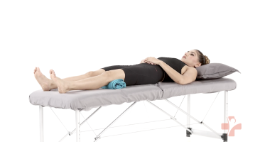 Quad sets (towel under knee)
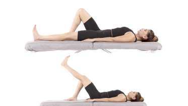 Straight leg raise
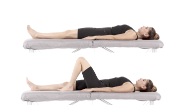 Heel slides
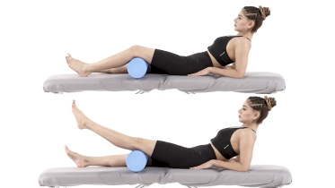 Short arc quads
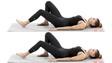 Hamstring sets (heel dig)
Stage 2: Build strength with weight on the leg
When you can walk without crutches and without a limp, with or without the brace, you can lift the leg straight without the knee sagging, and stage 1 feels easy. The band and the seated work strengthen the front and back of the thigh. The mini squat, the wall sit and the calf raises then train the leg with your weight on it. Bend only as deep as feels comfortable and under control, with your kneecap pointing over your second toe so the knee does not drift inward. Stand beside a kitchen counter for the single leg stance, so you can hold on if you need to.
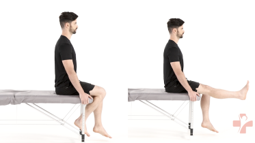 Seated knee extension
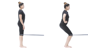 Terminal knee extension with band
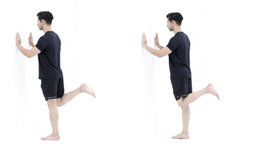 Standing hamstring curl
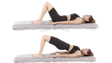 Glute bridge
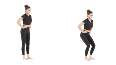 Mini squat
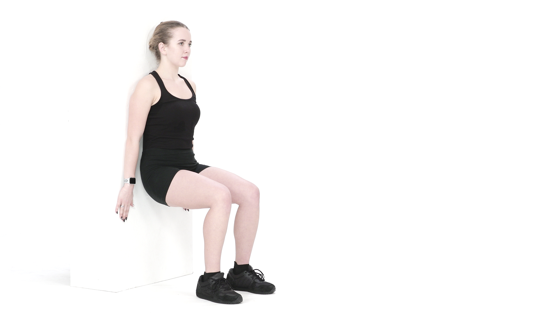 Wall sit
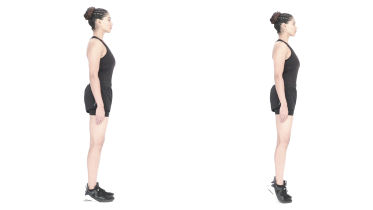 Calf raises
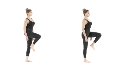 Single leg stance
Stage 3: Single leg strength and control
Once stage 2 feels easy and your knee is no worse the morning after. If you still wear a brace, ask your physio before you start this stage. Here one leg has to hold the knee steady while you step, lunge or reach, and that is where an inner knee that is still healing tends to wobble inward. Use a low step for the step up, and start the split squat and the lunge with a short range. In the single leg mini squat with band, the band pulls the knee inward, so add that exercise last in this stage, once the other exercises feel steady and the inner knee is no longer sore. Start with a light band and a gentle pull, and do the squat without the band if the inner knee hurts or the knee gives in to the pull. Side stepping is the first exercise that moves you sideways, so keep the steps small at first. For the clock reach, have a counter or wall close enough to grab.
 Step up
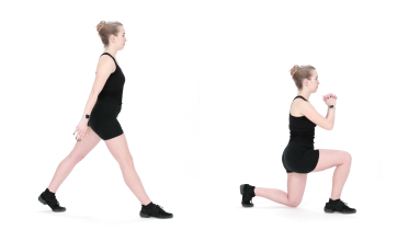 Split squat
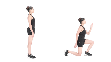 Reverse lunge
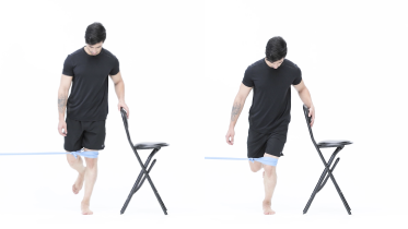 Single leg mini squat with band
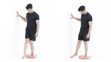 Single leg clock reach
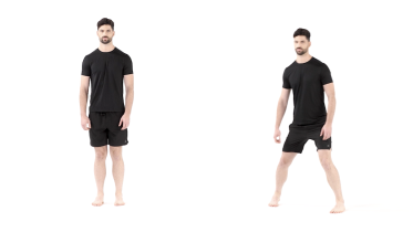 Side stepping
Stage 4: Jumping, hopping and the road back to sport
When single leg work feels solid and the inner knee no longer hurts or feels loose with it. Start with jumps on two feet, then hops on one, and only then the side lunge and skater hops, which push the knee sideways and load the healing ligament the most. If you have osteoporosis or have had a fall in the past year, ask your physio before you do any jumping or hopping. If your sport involves cutting and turning, your physio can add running drills that change direction and help you plan the final steps back to full training.
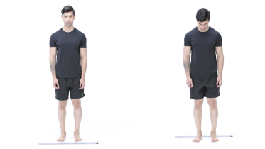 Line jumps
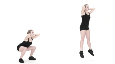 Jump squats
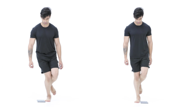 Single leg hops over a line
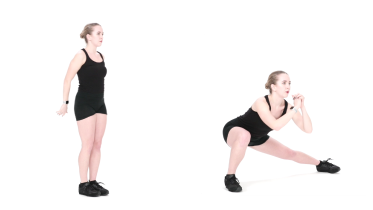 Lateral lunge
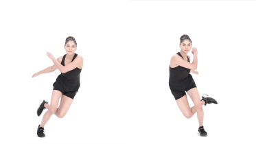 Skater hops
What to avoid while the ligament heals
Many physios hold back movements that push the knee inward or twist it until the ligament has had time to heal. In the early weeks that usually means no side lunges, cutting, pivoting or breaststroke kick, and no sport where someone might fall against the outside of your knee. This comes from common practice and from the way the injury happens. No trial has tested it. The program brings sideways movement back in stages 3 and 4, once the knee copes with straight line work.
Getting back to sport
The first question most people ask is how many weeks it will take. There is no firm answer. It depends on the grade of the sprain, on your sport and on how your knee responds, and the research gives no set return times for each grade.
Instead, many physios look for signs that the knee is ready. It moves fully, and the inner side is no longer tender or loose. Strength and hopping are close to the other leg. Running with changes of direction feels confident. These signs come from clinical practice and have not been tested as a set of rules.
OrthoInfo describes a gradual, stepped return to sport, and notes that a doctor may suggest a knee brace during sport to help prevent another injury. Kim and colleagues (2016) add that rehab should also deal with whatever made the injury likely, to lower the chance of it happening again.
When to see a physio or doctor
See a physio or doctor after any knee injury that leaves the inner knee painful, swollen or unstable. They can grade the sprain and pick up an ACL or meniscus injury early. Get help too if the knee is not improving as you expected, still feels loose weeks after the injury, or you are not sure which stage suits you. The warning signs further down say which problems need help sooner, from an emergency visit to an appointment within a few days.
If you have a heart or lung condition, or blood pressure that is not under control, check with your doctor before you start. If you have fallen recently or feel unsteady, keep a kitchen counter beside you for the standing exercises.
For physiotherapists
The program is a starting framework for patients with an isolated MCL sprain managed without surgery. Combined ACL and MCL injuries and postoperative rehab are outside its scope. Encinas-Ullán and Rodríguez-Merchán (2018) use the Fetto and Marshall grading: grade I without valgus laxity, grade II with valgus laxity at 30 degrees of flexion, and grade III with laxity at both 0 and 30 degrees. The same review cites nearly 78% of grade III injuries having an associated injury, 95% of those to the ACL.
Vosoughi and colleagues (2021) quote a side-to-side difference in medial gapping on valgus stress radiographs at 30 degrees of flexion of at least 3.2 mm as suggestive of a complete superficial MCL tear, and more than 9.8 mm as suggesting combined MCL and posteromedial corner injury. They advise early range of motion with weight bearing as tolerated, the brace locked in extension for walking until extension lag resolves, and closed chain work once full weight bearing is reached. Their surgical indications include a Stener-type lesion with the pes anserinus between the tibia and the ligament, laxity in full extension, anteromedial rotatory instability, suspected multiligament injury, valgus malalignment, an associated tibial plateau fracture and a chronic symptomatic MCL injury despite at least 6 weeks of non-surgical care. Encinas-Ullán and Rodríguez-Merchán state that isolated grade III tears without valgus instability can be managed non-surgically, although their own grading gives grade III laxity at 0 degrees. Read alongside the Vosoughi list, the page therefore treats laxity in full extension as a reason for surgical review rather than a route into this program.
The early functional protocol of Reider and colleagues (1994) used lateral hinged braces with unrestricted flexion and extension, early range of motion, then quad setting and leg raises, with resistance added at 90 degrees of flexion. Vosoughi and colleagues (2021) describe early range of motion as a key part of management to prevent arthrofibrosis. Kim and colleagues (2016) note that no level I studies compare rehab protocols, so the progression criteria here are pragmatic. Valgus and rotational loading come last, and stage 4 is matched to the athlete's sport.
See a doctor promptly if
- Emergency: the knee has moved out of place, looks misshapen or is pointing at an odd angle, or you have tingling or no feeling in the knee, leg or toes, or your foot turns cold, pale or blue. Go to an emergency department straight away. Do not drive yourself: ask someone to drive you or call an ambulance.
- Same day: the knee is locked, which means it is stuck bent and you cannot fully straighten it, even after gently trying to move it, or you cannot bend or straighten the leg at all. Get assessed the same day by a doctor, at urgent care or at an emergency department. A meniscus tear can happen together with an MCL injury, and a torn piece of meniscus can get caught in the joint and block it, so it needs checking early. A knee that catches for a moment and then frees itself can happen with a meniscus tear. That is not the same as a locked knee, but tell your physio or doctor about it.
- Emergency: the knee became very swollen very quickly, within a few hours of a twist, fall or blow, or you felt a pop or snap. Go to an emergency department straight away. Do not drive yourself: ask someone to drive you or call an ambulance. An MCL sprain on its own usually causes swelling over the inner side of the knee. When the whole knee swells within a few hours, it suggests bleeding inside the joint, which can come from a torn ACL, so fast swelling needs checking.
- Same day: you felt the kneecap slip out of place and it went back by itself. Get assessed the same day.
- Same day: the knee swells up quickly and a lot with no clear reason, especially if you take medicine to thin your blood. Get medical advice the same day.
- Same day: you cannot walk or put weight on the leg, or you have very bad knee pain after a fall or injury. Get medical advice the same day.
- Same day: the knee keeps giving way under you, or it gave way after a recent injury, or it gives way and is also very painful or swollen. Get medical advice the same day. If it only happens now and then, with no recent injury and no swelling, it is not an emergency, but book an assessment with a physio or doctor in the next few days.
- Same day: the knee is hot, red and swollen, or you have knee pain with a high temperature or feel hot, cold or shivery. Get medical advice the same day, and go to an emergency department if you feel very unwell. This can be a sign of infection in the joint, which needs treatment quickly.
- Same day: a calf or thigh that is newly swollen, warm, tender, red or darker than usual, has swollen veins that are sore to touch, or has a throbbing or cramping pain that does not fit your injury or condition, or that feels different from normal muscle soreness after exercise. This can be a sign of a blood clot (deep vein thrombosis), especially after surgery, an injury, time in a cast or boot, a long trip or a spell of being much less mobile than usual. Get medical advice the same day. If you are also short of breath or have chest pain, call emergency services.
- Within a few days: pain at night or at rest that keeps getting worse. See your doctor within a few days. If you have had cancer, now or in the past, see your doctor sooner, within a day or two, and mention it, even if the pain is not getting worse. Do the same if you have knee pain and are losing weight without trying, even if it does not hurt at night. If you are being treated for cancer now, contact your cancer team the same day.
Common questions
How long does an MCL sprain take to heal?
It depends mostly on the grade, and there is no fixed timeline. A mild (grade I) sprain may not need a brace at all, while one review says a hinged brace is usually worn for at least 3 weeks for a grade II tear and at least 6 weeks for a grade III tear (Vosoughi and colleagues, 2021). Getting back to sport takes longer than the brace time, because strength, hopping and turning have to come back too. Your physio will go by how the knee performs, not only by how many weeks have passed.
Can a torn MCL heal on its own?
Usually yes, and most people never need an operation. A review of isolated MCL tears says partial tears (grades I and II), and many complete (grade III) tears with no other ligament injury, can be treated without surgery through early rehab (Encinas-Ullán and Rodríguez-Merchán, 2018). Another review lists a knee that still opens up when pushed inward with the leg fully straight as one of the signs that point toward surgery (Vosoughi and colleagues, 2021), so a doctor checks for it. Healing without surgery does not mean doing nothing, though. The brace and the exercises are part of the treatment.
Can you walk with an MCL sprain?
Often yes, in a brace if you were given one. One review advises putting as much weight on the leg as it tolerates and starting knee movement as early as possible (Vosoughi and colleagues, 2021). Crutches help while walking still makes you limp. If you cannot put weight on the leg at all after the injury, get it checked the same day.
Do I need a knee brace for an MCL tear?
For a grade II or grade III tear, usually yes. The brace is hinged, so it lets the knee bend and straighten while it supports the inner side (Reider and colleagues, 1994; Vosoughi and colleagues, 2021). A grade I sprain can usually be managed without one. OrthoInfo notes that a doctor may also suggest a brace during sport after you return, to help prevent another injury.
Does a torn MCL need surgery?
Rarely on its own. Surgery is more likely when the MCL tear comes with other injuries, such as a torn ACL, when the ligament has pulled off a piece of bone or got trapped where it cannot heal, or when the knee stays unstable despite good non-surgical care (Encinas-Ullán and Rodríguez-Merchán, 2018; Vosoughi and colleagues, 2021). A knee surgeon makes that call after examining the knee.
References
- Encinas-Ullán CA, Rodríguez-Merchán EC. Isolated medial collateral ligament tears: an update on management. EFORT Open Reviews. 2018;3(7):398-407. https://doi.org/10.1302/2058-5241.3.170035
- Vosoughi F, Rezaei Dogahe R, Nuri A, Ayati Firoozabadi M, Mortazavi J. Medial collateral ligament injury of the knee: a review on current concept and management. Archives of Bone and Joint Surgery. 2021;9(3):255-262. https://doi.org/10.22038/abjs.2021.48458.2401
- Reider B, Sathy MR, Talkington J, Blyznak N, Kollias S. Treatment of isolated medial collateral ligament injuries in athletes with early functional rehabilitation. A five-year follow-up study. American Journal of Sports Medicine. 1994;22(4):470-477. https://doi.org/10.1177/036354659402200406
- Kim C, Chasse PM, Taylor DC. Return to play after medial collateral ligament injury. Clinics in Sports Medicine. 2016;35(4):679-696. https://doi.org/10.1016/j.csm.2016.05.011
- NHS. Knee pain. Page last reviewed 21 December 2023. https://www.nhs.uk/symptoms/knee-pain/
- NHS. Deep vein thrombosis (DVT). Page last reviewed 30 April 2026. https://www.nhs.uk/conditions/deep-vein-thrombosis-dvt/
- American Academy of Orthopaedic Surgeons. Collateral Ligament Injuries. OrthoInfo. https://www.orthoinfo.org/en/diseases--conditions/collateral-ligament-injuries/
Written and checked by the PocketPhysio editorial team. Last updated 2026-09-28.
This page is general education, not a diagnosis or a personal treatment plan. See a physiotherapist or doctor for advice about your own situation.