
PCL injury exercises and recovery by stage
Warning signs, and when to get help
What is a PCL injury?
The posterior cruciate ligament is the thicker of the two ligaments that cross in the middle of your knee. Its job is simple to describe: it stops the shin bone sliding backward under the thigh bone. When it is sprained or torn, the shin can drop back a little, most noticeably when the knee is bent.
A PCL injury is graded by how far the shin shifts on examination. Grades I and II are partial, grade III is a complete tear. The grade is useful but it does not run your life: in the largest natural history study of these injuries, how loose the knee was did not predict how well it worked or how people scored years later (Shelbourne and colleagues, 1999).
This page is for adults with an injury that involves the PCL on its own, managed without an operation, including people still deciding. If more than one ligament was torn, or your knee was reconstructed, the plan comes from your surgeon and your physio instead.
How a PCL gets injured, and what it feels like
Two mechanisms do most of the damage. A blow to the front of the shin while the knee is bent, which is the classic dashboard injury in a car crash and the fall onto a bent knee in sport. And a forced straightening or twist that overloads the back of the joint.
What people describe afterward is often oddly undramatic. An ache at the back of the knee. Pain on kneeling. Swelling that comes up over hours rather than within minutes. A knee that feels unreliable going downstairs or slowing from a run, without ever quite giving way.
Plenty of people never heard a pop and did not think the injury was serious at the time, which is why some PCL injuries are found months later.
The danger is not usually the PCL itself. It is what else happened in the same moment. Enough force to drive the shin backward can tear the collateral ligaments, the ACL and the meniscus with it, and in the worst version the whole knee dislocates and the artery or the nerve behind it is damaged. Medina and colleagues (2014) reviewed that complication across the published series, and Ockuly and colleagues (2020) set out how these knees are assessed on the day. The knee can look close to normal afterward, which is exactly why the first warning signs on this page send you to hospital rather than to an examination couch.
Does a PCL injury need surgery?
For an isolated injury, usually not, and the medium-term results are good enough to be reassuring. Patel and colleagues (2007) followed 57 patients with acute, isolated, partial or complete PCL injuries treated without surgery for an average of 6.9 years: 53 of the 58 knees scored good or excellent on a knee function score, and the amount of looseness did not correlate with the score. Shelbourne and colleagues (1999) followed 133 athletically active people, average age 25, for an average of 5.4 years and found the same independence between laxity and function.
Grassmayr and colleagues (2008) pooled 47 articles, 17 of them on non-operative outcomes, and concluded that most grade I and II isolated injuries treated without surgery returned to activity. They also found the evidence for how the muscles compensate to be lacking, which is worth noting, because the idea that the quadriceps simply take over for a torn PCL is stated far more confidently online than the research supports. What the laboratory work does support is narrower and still useful. Quadriceps pull reduces the strain in the PCL between 20 and 60 degrees of knee bend (Dürselen and colleagues, 1995), which is a good reason to train the thigh and to train it in that range. It is not evidence that a strong thigh replaces the ligament, and nobody has shown that the muscles take over the ligament's job.
Surgery becomes the conversation when the PCL is torn along with other ligaments, when the knee is unstable enough to interfere with life despite good rehabilitation, or when a piece of bone has been pulled off with the ligament. That decision belongs with a knee surgeon who has examined you and seen your scans.
A 2025 Delphi statement from the German Knee Society describes the same shape. Non-operative treatment is the first approach for an acute isolated PCL injury, because the ligament has some capacity to heal, while reconstruction is usually indicated for a young, active person with high-grade instability (Eberle and colleagues, 2026). Two points from it matter if your injury pulled a piece of bone off the back of the shin along with the ligament. Reading the published series, that panel put surgical reduction at displacement of more than about 5 to 6 mm, and every member of it agreed on repeat X-rays from roughly two to three weeks after injury when this kind of injury is being treated in a brace, so that a fragment slipping is caught while the plan can still be changed. If that is your injury, keep those follow-up appointments.
PCL injury recovery time: what the studies actually found
Only one of the studies above gives return-to-sport timings, and it studied athletes. Agolley and colleagues (2017) followed 46 athletes with isolated grade II or III PCL injuries, seen within four weeks of injury and treated with bracing and then individualised rehabilitation. Return to sport-specific training averaged 10.6 weeks, and return to full competitive sport averaged 16.4 weeks, with a range of 10 to 40 weeks. At two years, 42 of the 46 were playing at the same level or higher; at five years, 32 were.
Two things to take from that. The spread of 10 to 40 weeks is the honest answer to how long this takes, and the five-year figure is lower than the two-year one, which tells you that getting back is not the same as staying back.
The larger natural history series is less rosy about sport, and it belongs next to those figures. Among the 133 athletically active patients Shelbourne and colleagues (1999) followed, and whatever the grade of looseness, half went back to the same sport at the same level or higher, a third went back at a lower level, and a sixth did not go back to it at all. Agolley's athletes were all treated within four weeks of the injury by one team that braced them first, which is not everybody's starting point.
Searches for PCL recovery time return football and NFL return dates. Those are reported by journalists about professional athletes with daily medical supervision, surgical options, and reasons to return that have nothing to do with their knee. They are not evidence about yours, and nothing on this page is built on them.
Why PCL rehab is not ACL rehab turned around
The ligament fails in the opposite direction, so the exercises that are safe early are the opposite too, and this is the part most generic knee programs get wrong.
The hamstrings pull the shin backward. That is precisely the movement the injured PCL is supposed to resist, so isolated hamstring curls, especially lying on your front or sitting against resistance, are kept out of the early weeks. In their place, the quadriceps do the work: the published rehabilitation review recommends progressive weight bearing, preventing the shin from sliding backward, and strengthening the quadriceps (Pierce and colleagues, 2013).
Laboratory work puts angles on this. Dürselen and colleagues (1995) loaded nine cadaver knees in a rig that let the joint move freely, simulated pull from the thigh, hamstring and calf muscles, and measured the strain in the cruciate ligaments. Hamstring pull strained the PCL between 70 and 110 degrees of knee bend. Quadriceps pull did the opposite between 20 and 60 degrees: it reduced PCL strain. A strong calf contraction strained the PCL at anything beyond 40 degrees of bend, which is why those authors advised against strong calf work past 30 degrees of knee bend after PCL surgery.
Nine cadaver knees are not a group of patients, so take those angles as the reasoning behind the plan rather than as limits for your own knee. What they come to in practice is simple. The thigh work sits in the range that helps, resisted knee bending waits, and the heel raises in stage 2 are done standing with the knee straight, not seated or with the knee bent.
Bending the knee needs thought too. Sitting or lying on your back with the knee bent lets gravity pull the shin backward, so the same review recommends bending the knee lying face down, where the thigh is supported and the shin's own weight does not drag it the wrong way. That is why prone knee bending is the first bending exercise in stage 2.
Read that recommendation closely, though, because it is for passive movement. The exercise page teaches the prone knee bend as an active lift, worked by the back of the thigh, and the back of the thigh is the pull this ligament resists. Draw the heel up with a towel or a belt around the ankle instead, with an end in each hand up by your shoulders, and keep the hamstrings relaxed. It is the same movement without the contraction.
Weight bearing is the friendlier direction. With the foot planted, the thigh muscles and the joint surfaces share the load, and the shin is not left hanging. So squats, wall sits, step ups and split squats arrive earlier in this program than you might expect, and seated leg curls do not arrive at all.
Depth is the part to hold back. Load on this ligament climbs as the knee bends further, and the range in which the thigh muscles protect it runs out somewhere around 60 degrees, so there is a single ceiling on every loaded knee bend in this program rather than a range to aim at: stop while your hips are still clearly higher than your knees, which is roughly 60 degrees of bend and well above the right angle the wall sit video shows. We give you the landmark rather than the number because you cannot measure a knee angle at home, and a reader given a range reads the deep end of it as the target. If you are unsure, go less far rather than further. Deepening it is a decision for the physio or surgeon who examined your knee, not something to judge by how it feels on the day, because the load rises with depth whether or not it hurts.
How to use this program
Start at stage 1 if the injury is recent or the knee is still swollen. If it happened a while ago and the knee is only unreliable under load, stage 2 or 3 may fit, and a physio can tell you which. Move up when the current stage is easy and the knee is no worse the day after.
Each exercise page gives a typical starting dose. Across this program that is mostly 2 to 3 sets of 8 to 15 in the first stages and 2 to 3 sets of 6 to 12 for the single leg work later, with holds at 5 to 10 seconds and the wall sit at 10 to 30 seconds. The lying and sitting work can be done daily, and the harder standing strength work 2 or 3 days a week so the muscles recover. Your physio will adjust the exercises and the numbers to your knee and your sport.
One rule runs through every stage. Wherever the knee bends under load, the depth stays capped until a clinician lifts it, and that applies to the wall sit, the squat, the split squat, the lunge and the step down alike. The cap is one ceiling and not a range: stop while your hips are still clearly higher than your knees, which is around 60 degrees of knee bend. The exercise pages are written for knees in general and several of them teach a right angle or deeper, with the thighs level with the floor, so the cap on this page is the one to follow and the depth in the videos is not.
Before you start, two checks. Has the knee been examined for other ligament damage, because an isolated PCL injury and a multi-ligament injury are not the same problem. And were you given a brace, because if you were, this program runs inside your clinician's instructions for it rather than around them. Ask your physio before adding any resisted hamstring work.
If you have had knee surgery of any kind, a repair, a reconstruction, a cartilage trim or a replacement, your surgeon's or hospital team's own instructions come first, including where they differ from anything on this page. Anything new in an operated knee goes to that team, not to a web page. New pain, swelling, heat or giving way in a knee that has been operated on is a same-day call to them, for the same reason the warning list sends a hot, red and swollen knee for advice the same day: an infection, or a problem with the repair or the implant, has to be ruled out before anyone treats it as a strain.
If your PCL was reconstructed, the numbers that matter are your surgeon's, and the published picture shows why this page will not guess them. When the rehabilitation protocols used after isolated PCL reconstruction were collected from 120 orthopaedic institutions in Germany, Austria and Switzerland, only 33 had a protocol at all and no two of those were identical: in the first week after surgery some allowed 10 degrees of knee bend and others 60, one allowed whatever was comfortable, and the weight permitted on the leg ranged from none to all of it (Memmel and colleagues, 2022). What did hold across them was the direction of travel. More than 90% kept weight bearing at half body weight or less and knee bend at 60 degrees or less for at least the first two weeks, and every protocol that mentioned a brace required one. So after a reconstruction, work to your own team's weight bearing and bending limits rather than to the stages below, and ask them before you move up.
Breathe normally through every repetition rather than holding your breath, especially if you have high blood pressure. Check with your doctor before you start if you have a heart or lung condition, or blood pressure that is not under control. If you have fallen in the past year or feel unsteady on your feet, keep a hand on a kitchen counter through the standing exercises. If you are pregnant, the exercises done lying on your front or flat on your back need changing as pregnancy goes on, so plan those with your physio and your maternity team.
Read the warning signs on this page before you start, not after something happens. They sit below the program because that is how every program page here is laid out, and the three at the top of that list are the ones that mean hospital rather than a lighter session: a knee that felt as though the whole joint shifted out of place at the moment of injury, a knee that swelled up within a few hours, and numbness, tingling or a foot that turns cold, pale or blue. Two more are worth carrying in your head while you work through the stages. A calf or thigh that becomes swollen, warm, tender or sore to touch needs advice the same day, because a blood clot is one of the causes and a spell of being much less mobile makes it likelier. And a knee that gives way under you means leaving out the exercises you do standing on one leg until it has been checked.
The exercise program
Stage 1: Protect the knee and wake up the thigh
For the first weeks, and while any brace your clinician gave you is in use. Everything here is done lying or sitting, and the aim is to get the quadriceps working again without letting the shin drop backward. That is why there is no hamstring exercise in this stage: the hamstrings pull the shin in exactly the direction the injured ligament is meant to stop. Quad sets and short arc quads work the last part of straightening, which is the part a swollen knee gives up first. Kneecap glides keep the kneecap moving, and the ankle pumps keep the calf working while you are less mobile. Many programs use 2 to 3 sets of 10 to 15 for these, once or twice a day, with the holds at 5 to 10 seconds, and your physio will adjust this. Keep this stage pain free.
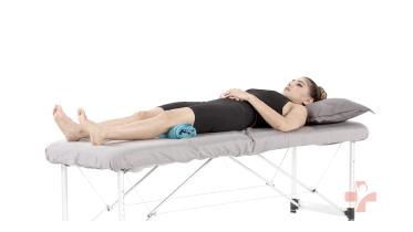 Quad sets (towel under knee)
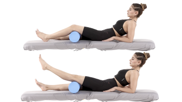 Short arc quads
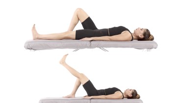 Straight leg raise
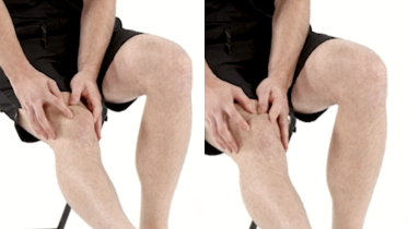 Kneecap glides
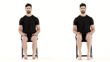 Ankle pumps
Stage 2: Get the bend back, with the shin supported
Start when stage 1 is easy and your physio or surgeon agrees. Bending the knee is where a PCL injury needs care, because gravity pulls the shin backward when you bend the knee sitting or lying on your back. The published rehabilitation review recommends bending the knee lying face down for that reason: the thigh rests on the bed and the shin is supported by the leg's own weight rather than hanging (Pierce and colleagues, 2013). So prone knee bending comes first here, with one change that matters for this ligament. What that review recommends is passive bending, so let the knee be drawn into the bend rather than pulling it there with the back of the thigh. Set it up like this: lying on your front, loop a towel, a belt or a dressing gown cord around that ankle, bring the two ends up over your shoulders so one is in each hand, and pull down gently with your arms so the heel travels toward your bottom. Your arms do the work and the back of the thigh stays soft. Lower the foot the same way, letting the towel out rather than dropping the leg. Stop where it feels like a stretch rather than going for more bend. If you cannot set that up, ask your physio for another way to bend the knee rather than doing the active version in the video. The heel prop works on getting the knee fully straight, and it stops at straight: let the knee sink down to level and no further, and if your knee already bends backward past straight, leave this one out and ask your physio, because a forced straightening is one of the two ways this ligament gets injured. Then the standing work begins: terminal knee extension, a shallow squat, a wall sit and heel raises all load the knee with your foot on the floor, which is the safer direction for this ligament. One setup detail in the terminal knee extension: sit the band just below the crease at the back of the knee, across the top of the calf, rather than up on the back of the thigh. A band behind the thigh pulls the thigh bone forward over a planted shin, which is the same relative movement this ligament is there to stop, and a band on the calf pulls the other way. It costs you nothing to place it a few centimetres lower. Expect 2 to 3 sets of 8 to 15, with holds of 10 to 30 seconds for the wall sit. Keep the wall sit and the squat shallow, and use one ceiling rather than a range: stop while your hips are still clearly higher than your knees. That is roughly 60 degrees of knee bend, and it is well above the right angle with the thighs level with the floor that the wall sit page describes. Nobody can judge degrees at home, so if you are unsure, go less far rather than further, because the load on this ligament climbs as the knee bends and less bend is never the risky choice. Do the heel raises standing with the knee straight rather than seated or with the knee bent. Ask your physio before you do seated knee bends against resistance.
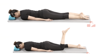 Prone knee bend
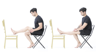 Seated heel prop
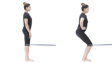 Terminal knee extension with band
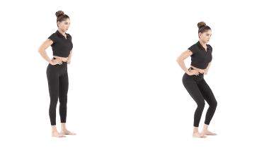 Mini squat
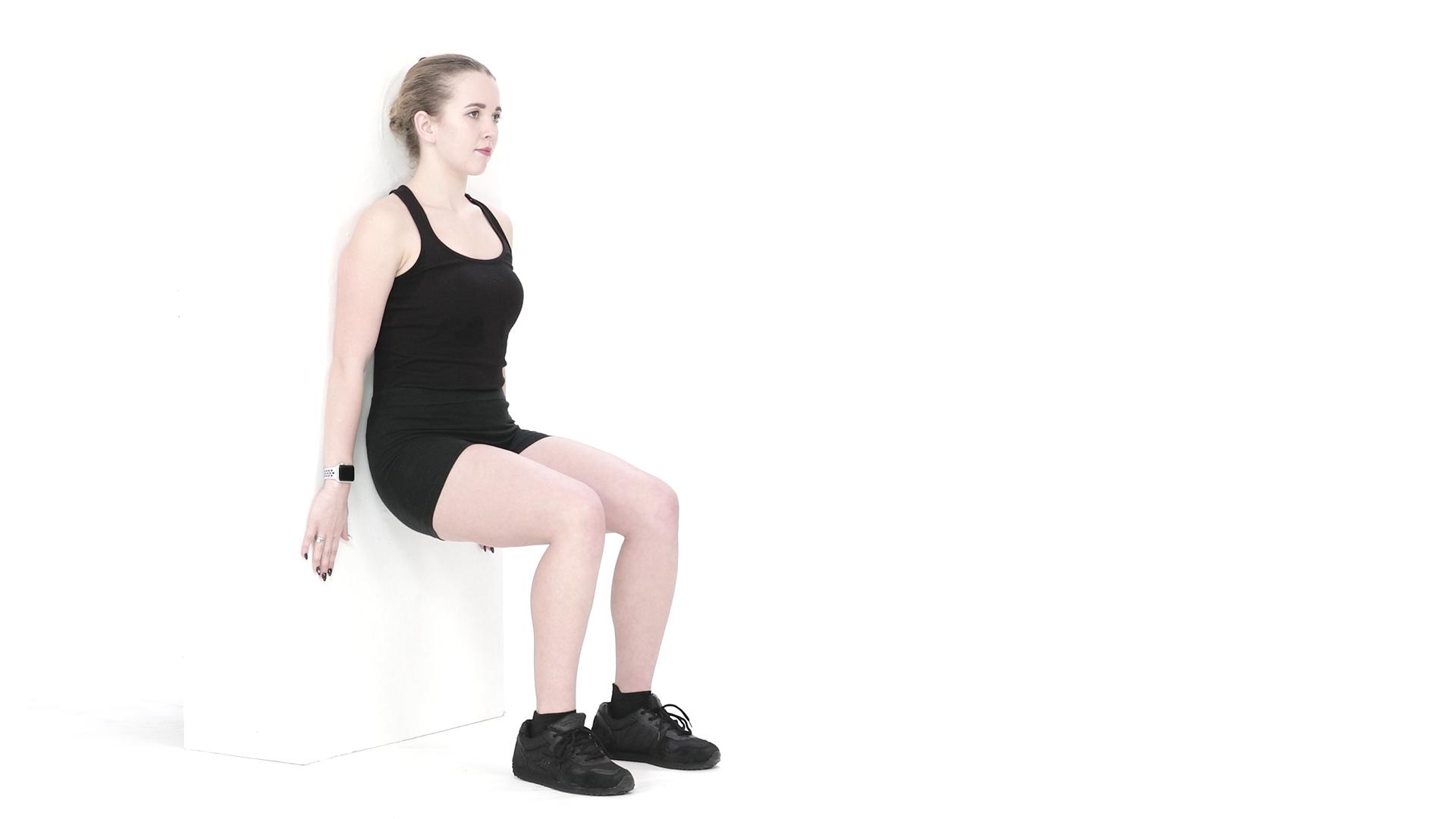 Wall sit
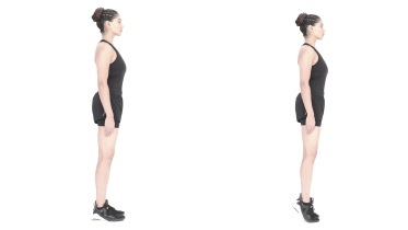 Calf raises
Stage 3: Build real strength in standing
Move up when stage 2 feels easy and the knee settles well afterward. These train the knee under load with the foot planted, which is what stairs, hills and getting out of a car ask for. Start the squat shallow and deepen it only as far as your physio has cleared. The same ceiling as stage 2 applies: stop while your hips are still clearly higher than your knees. A chair behind you to touch down on helps you find the same depth every time, but build the seat up with firm cushions first, because touching a normal chair seat takes the knee to about a right angle, which is past the cap. Comfort is not the limit for this ligament, because the load on it rises with depth whether or not the knee complains. That cap covers everything here that bends the knee under load, and not the squat alone, so keep the split squat shallow as well and use a low step, about the height of one stair, for the step up. The exercise pages describe deeper versions, a squat down to where the thighs are level with the floor and a split squat with the back knee near the floor, and those wait until a clinician lifts the limit. The step up practices stairs, the split squat loads one leg at a time, and prone hip extension builds the buttock muscle without pulling the shin backward, as long as that knee stays straight through the whole lift. Let the knee bend and the same exercise turns into hamstring work, which is the pull this ligament resists, and a bending knee is exactly what the leg drifts into once the buttock tires. Usually 2 to 3 sets of 8 to 12, 2 or 3 days a week, so the muscles recover in between. If you have high blood pressure, breathe steadily through every repetition and never hold your breath. Mild discomfort that settles by the next morning is fine; a knee that is sorer or more swollen the next day means you did too much.
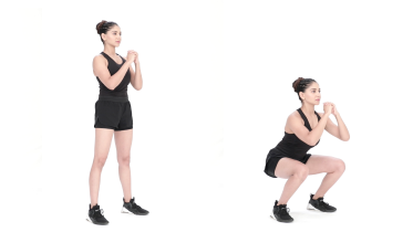 Squat
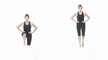 Step up
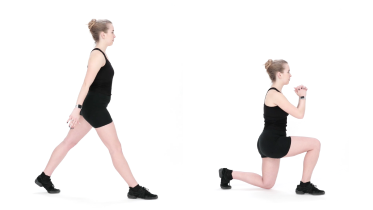 Split squat
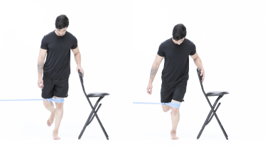 Single leg mini squat with band
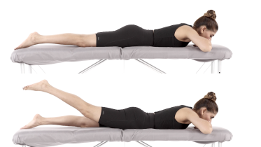 Prone hip extension
Stage 4: One leg, direction changes and back to sport
This is the stage that decides whether the knee copes with sport, and it is the one most people stop short of. Every exercise loads one leg, three of them with the knee bending under control, which is the demand a step down, a landing and a change of direction all make. In the single leg deadlift the knee stays almost straight and the movement happens at the hip, which is why it is here rather than in stage 1. Keep a counter close for the balance and step down work at first. In the single leg stance all the work is in the leg you are standing on, so keep the lifted leg fairly straight, or hold the thigh up with the foot forward, rather than letting the shin hang down at a right angle as the exercise page shows. A hanging shin is being pulled backward by its own weight at the knee, which is the same reason this page tells you not to let the lower leg dangle when you sit. The step down, the lateral lunge and the single leg squat all bend the knee under load, so they stay inside the depth your physio has cleared, under the same ceiling as the earlier stages: hips clearly higher than knees. A low step comes before a higher one, and the step height is what sets the depth here, so change it in small amounts rather than moving straight to a full stair. Normal practice is 2 to 3 sets of 6 to 12 on each side, 2 or 3 days a week. Running, cutting and jumping come after this stage, built up on a plan you agree with your physio, and a knee that still feels as though it could shift under you is not ready for them.
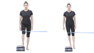 Lateral step-down with band
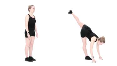 Single leg deadlift
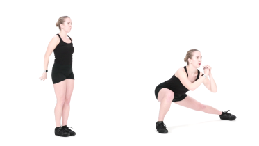 Lateral lunge
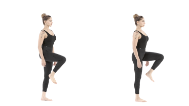 Single leg stance
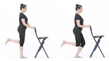 Supported single leg squat
Kneeling, stairs and the things that actually bother people
Kneeling is the complaint we hear most, and it rarely gets a mention in the research. The front of the shin presses into the floor in exactly the direction the ligament resists, so it can stay sore long after walking is comfortable. That same direction is the reason kneeling is left out early rather than padded: in the first weeks, and for as long as you are in a brace for this injury, treat it as something to avoid and ask your physio when it can start, not as a comfort problem to solve with a cushion. Later, a thick pad, kneeling on one knee rather than both, and getting up every few minutes make most jobs possible. Expect it to be one of the last things to come back.
One small habit helps all day. When you sit with the knee bent, rest the foot on the floor or a low stool rather than letting the lower leg dangle, because a shin with nothing under it is being pulled backward for as long as it hangs there.
Going downstairs is the other one. Lowering your body weight with the knee bent is the position in which the PCL is working hardest, so stairs and slopes often feel unreliable before anything else does. Holding the rail and taking one step at a time is sensible in the first weeks, and the step down work in stage 4 is what gets you off it.
The compensation to watch for is the same one every sore knee produces: a quiet shift of weight onto the other leg all day long, standing on it while the kettle boils and leaning over it to get out of a chair. None of it is wrong and all of it keeps the injured side weak.
What we notice in the clinic
The thigh is the story. A knee with a PCL injury loses quadriceps size and strength quickly and gets it back slowly, and almost everyone who is still struggling at six months has a thigh that is visibly smaller on that side. Measuring it, rather than guessing, changes what people do.
Then there is the flat ground. The knee often feels fine walking on the flat quite early, which gives a false sense of recovery. Slowing down, going downhill and stopping suddenly are the tests that find the deficit, and they are the ones people avoid without noticing.
And the hamstring question, which comes up every time. People who lift weights want to know when they can do leg curls again, and the answer is that it waits, it comes back through hip-dominant work first, and it is a question for the physio who knows how your knee examines rather than for a page.
When to see a physio or doctor
See someone if a knee injury left you with pain behind the knee, swelling, or a knee you do not trust, even if you can walk on it. A PCL injury that nobody has examined is easy to mistake for a strain, and the examination is quick.
Go sooner if the knee gives way, if you cannot fully straighten it, or if you are not making progress after several weeks of regular work. Ask for a reassessment before you go back to a sport with cutting and pivoting, and ask for your thigh strength to be measured rather than estimated.
The warning signs below say what needs help faster, and how fast. The first three are there because a high-energy knee injury can involve far more than the PCL, and some of that damage is not obvious on the day.
For physiotherapists
The program follows the principles in Pierce and colleagues (2013): progressive weight bearing, prevention of posterior tibial subluxation, quadriceps emphasis, and prone passive flexion so the tibia is not loaded posteriorly by gravity. That is a Level IV review that proposes guidelines rather than testing them, and the page says so. Open-chain knee flexion is deferred and the patient is told to ask before adding it.
The page now states the angles rather than only the mechanism, traced to the study that produced them: in nine cadaver knees with simulated muscle loading, hamstring activation strained the PCL from 70 to 110 degrees of flexion, quadriceps activation reduced PCL strain from 20 to 60 degrees, and gastrocnemius activation strained it beyond 40 degrees, on which the authors recommended avoiding strong gastrocnemius contraction past 30 degrees of flexion after PCL surgery (Dürselen and colleagues, 1995). Those numbers are labelled as cadaveric on the page and used to justify the shallow cap on the wall sit and the squat, the straight-knee heel raise, and the exclusion of seated or bent-knee calf work, rather than as patient limits. No figure from the protocol in circulation for unsupported flexion is used, because that document could not be read here; where a number appears it comes from Dürselen or from the protocol survey below.
Four technique points are carried on this page rather than on the library exercise pages, which are written for knees in general. Pierce and colleagues recommend prone PASSIVE flexion, and content/exercises/prone-knee-bend.md teaches active prone flexion past the vertical with the hamstrings named as the working muscle, which is a loaded contraction inside the 70 to 110 degree arc. This page therefore instructs a towel or belt assisted bend with the hamstrings relaxed, and now says how the strap is held, an end in each hand up by the shoulders, because an instruction a prone patient cannot physically carry out is an instruction to do the version in the video instead. The depth cap is likewise stated for every loaded knee bend in the program, rather than for the wall sit and the squat alone, because content/exercises/split-squat.md offers a full version with the back knee near the floor and content/exercises/squat.md takes the thighs to level with the floor. The mini squat (30 to 40 degrees), supported single leg squat, single leg mini squat with band and lateral step down with band are all written at or below the cap already and needed no restriction.
The cap is now written as one ceiling with an observable landmark rather than as a 45 to 60 degree range, because a range is read as a target at its deep end and a home exerciser cannot estimate either number: hips clearly higher than knees, which also distinguishes the capped position from the thighs-level-with-the-floor depth the wall sit and squat videos show. Nothing was permitted that the range did not already permit. The two remaining cues are mechanical rather than trial based and are labelled as such: content/exercises/terminal-knee-extension.md places the band "behind that knee" without specifying above or below the joint line, and a band anchored anteriorly across the distal femur produces relative posterior tibial translation, which is the one thing Pierce and colleagues' protocol exists to prevent, so the page asks for the loop across the proximal calf. And content/exercises/seated-heel-prop.md is a multi-minute gravity-loaded extension stretch with no end point other than straight, prescribed on a page that names hyperextension as one of two injury mechanisms, so the page now stops it at neutral and routes a knee that already hyperextends to its physio.
Post-operative limits are not set by this page and are attributed with their variation. Of 120 orthopaedic institutions in German-speaking countries, 33 had a protocol after isolated PCL reconstruction and none were identical, with first-week flexion caps of 0 to 10 degrees in 35.5%, up to 30 degrees in 45.2% and up to 60 degrees in 16.1%, while more than 90% held weight bearing at half body weight or less and flexion at 60 degrees or less for at least the first two weeks (Memmel and colleagues, 2022). The bracing figures for conservative care, six weeks static then six weeks dynamic and not less than three months in total, are from a Delphi expert statement graded C and E with 80% agreement, and are presented at that strength (Eberle and colleagues, 2026). Both are on the page because a reader who has had a reconstruction will arrive here, and the page routes them to their own team's numbers.
One in vivo finding supports the clinic note about level walking. Dynamic stereo radiography of nine patients with asymptomatic isolated grade II PCL deficiency found about 2 mm of posterior tibial subluxation during the swing phase of running with no stance phase difference, and about 4 mm during terminal swing and early stance of stair ascent with anterior reduction velocities 3 to 4 times the intact side (Goyal and colleagues, 2012). Level walking showed no difference, but it was evaluated in only 3 of the 9 patients and not tested in the remaining 6, so that is a negative finding in three knees rather than in nine. The measured abnormality is in stair ascent, while the symptom patients report is descending, so the page keeps both the level-walking note and the stair symptom as clinical experience and claims the kinematics for neither.
Outcome figures are attributed with their populations, under the rule that a number is traced to the paper that produced it. Shelbourne and colleagues (1999): 133 athletically active patients, mean age 25.2, acute isolated nonoperative PCL injuries, mean 5.4 years of subjective follow-up with 68 returning for clinical review, no change in laxity, and function independent of laxity grade. Patel and colleagues (2007): 57 patients, 58 knees, acute isolated partial or complete injuries, mean 6.9 years, Lysholm-II mean 85.2, 40% excellent and 52% good. Agolley and colleagues (2017): 46 athletes, 25 Hughston grade II and 21 grade III, bracing then individualised rehabilitation, return to sport-specific training at a mean of 10.6 weeks and to full competitive sport at a mean of 16.4 weeks with a 10 to 40 week range, 91.3% at the same level or higher at two years and 69.5% at five.
Two claims that recur in the ranking material are handled rather than inherited. The first is that the quadriceps functionally substitute for the PCL: Grassmayr and colleagues (2008) found the evidence for compensatory muscle activity lacking, so the page presents quadriceps work as the recommended emphasis rather than as a substitution mechanism. The second is a high rate of medial and patellofemoral arthrosis after conservative care: the contact-pressure basis for that claim is in Grassmayr, and it is reported as something that may cause articular damage, while the conservative cohorts show milder radiographic change at medium term, with 7 mild and 3 moderate medial and 4 mild patellofemoral in 58 knees at a mean 6.9 years (Patel and colleagues, 2007) and no correlation between joint space narrowing and laxity (Shelbourne and colleagues, 1999). Two papers reached in this review complete it and are now on the page: 44 of 68 nonoperatively treated acute isolated injuries examined at a mean 14.3 years, range 10 to 21, with radiographs normal or nearly normal in 39, moderate to severe osteoarthritis in 11%, and no difference by laxity grade (Shelbourne and colleagues, 2013); and a population-based incidence cohort of 48 isolated PCL tears matched to uninjured controls, with a hazard ratio of 6.2 (95% confidence interval 1.8 to 21.2) for symptomatic arthritis over a mean 12.2 years, non-significant hazard ratios for meniscal tear and arthroplasty, and older age at injury associated with both arthritis and arthroplasty (Sanders and colleagues, 2017). The page states the modest absolute rates, the significant relative risk and the absence of follow-up into old age, in that order, so the requalification no longer reads as reassurance.
Screen every acute PCL injury for a multi-ligament pattern and for a reduced knee dislocation. Vascular and neurological injury rates after knee dislocation are reviewed in Medina and colleagues (2014), and the acute evaluation and classification in Ockuly and colleagues (2020); the emergency warning signs on this page route a knee that shifted out of place, multidirectional laxity, rapid haemarthrosis and distal neurovascular change to an emergency department on the day.
Warning signs
Get emergency help now
- Emergency: the knee has moved out of place, looks misshapen or is pointing at an odd angle, or you have tingling or no feeling in the knee, leg or toes, or your foot turns cold, pale or blue. Go to an emergency department straight away. Do not drive yourself: ask someone to drive you or call an ambulance.
- Emergency: the whole knee, not just the kneecap, felt as though it slid or shifted right out of place at the moment of injury, even if it went back straight away, or the knee now feels loose or wobbly in more than one direction. Go to an emergency department straight away. Do not drive yourself: ask someone to drive you or call an ambulance. A knee that comes out of place and goes back can tear several ligaments at the same time and can damage the artery or the nerve behind the knee. That damage is not always obvious at first, and the knee can look almost normal, so it is checked in hospital on the day rather than worked out at home.
- Emergency: the knee became very swollen very quickly, within a few hours of a twist, fall or blow, or you felt a pop or snap. Go to an emergency department straight away. Do not drive yourself: ask someone to drive you or call an ambulance.
Get help today
- Same day: the knee is locked, which means it is stuck bent and you cannot fully straighten it, even after gently trying to move it, or you cannot bend or straighten the leg at all. Get assessed the same day by a doctor, at urgent care or at an emergency department. A knee that catches for a moment and then frees itself is not the same as a locked knee, but tell your physio or doctor about it.
- Same day: the knee straightens when someone else moves it for you, but you cannot lift the straight leg off the bed yourself or hold the knee straight against gravity, or you can feel a gap or a dip just above or just below the kneecap. Get assessed the same day by a doctor, at urgent care or at an emergency department. The tendon that straightens the knee can tear, which is not a ligament problem and is not treated with exercises. It is usually repaired, and a tendon repaired late does less well than one repaired early.
- Same day: you cannot walk or put weight on the leg, whether or not there was a fall or injury, or you have very bad knee pain after a fall or injury, or very bad knee pain that came on suddenly with no injury at all. Get medical advice the same day.
- Same day: the knee keeps giving way under you, or it gave way after a recent injury, or it gives way and is also very painful or swollen. Get medical advice the same day. If it only happens now and then, with no recent injury and no swelling, it is not an emergency, but book an assessment with a physio or doctor in the next few days. Until it has been checked, leave out the exercises you do standing on one leg, hold the rail on stairs and take care on uneven ground, because a knee that gives way without warning is how people fall.
- Same day: since your knee ligament was reconstructed, your knee gives way, or you feel a pop, a shift or a twist inside it, and it swells up within a few hours or is very painful. Keep your weight off it and contact your surgical team, or get assessed the same day at urgent care or an emergency department. A fall or a twist can damage the new ligament (graft) or a meniscus repair. If the knee looks out of shape, or your foot turns numb, cold or pale, go to an emergency department straight away, and do not drive yourself. One route sits above that one: if the knee swelled very quickly after a pop or a snap, the emergency department line in the warning signs above applies whichever operation you had, and a call to your surgical team is not a substitute for it. If the knee only feels a little unsteady now and then, with no swelling or pain, it is not an emergency, but tell your physio or surgeon in the next few days. After a posterior cruciate ligament reconstruction there is more to watch for than a single bad moment. If the knee swells up much more after a kneel, a deep bend, a twist or simply doing more than your plan allows, even with no pop and nothing giving way, stop the exercises, keep your weight off it, go back to your brace and crutches if you were given them, and contact your surgical team the same day. This graft also stretches out quietly more often than it fails in one go, so a knee that feels looser week by week, a shin that feels as though it sits further back, or kneeling, squatting and stairs getting harder rather than easier, is worth a call to your surgical team rather than waiting for the next appointment, even though none of that is an emergency.
- Same day: since your operation, your wound is oozing, leaking or has pus coming out of it, or its edges start to come apart, or your knee is tender, swollen, painful or red and it is not getting better or is getting worse, or you have a high temperature or feel hot and cold or shivery. Redness can be harder to see on brown or black skin. Contact your surgical team the same day, or get urgent medical advice if you cannot reach them. These can be signs of an infection in the wound or the knee joint. If you feel very unwell, go to an emergency department. If you have a very high or very low temperature, shivering you cannot control, you are breathing very fast or finding it hard to breathe, you are confused or your speech is slurred, your skin, lips or tongue look blue, gray, pale or blotchy (on brown or black skin this can be easier to see on the palms of the hands or soles of the feet), or you have a rash that does not fade when you press it, call emergency services, as this can be sepsis.
- Same day: the knee is hot, red and swollen, or you have knee pain with a high temperature or feel hot, cold or shivery. Get medical advice the same day, and go to an emergency department if you feel very unwell. This can be a sign of infection in the joint, which needs treatment quickly.
- Same day: the knee swells up with no clear reason, especially if you take medicine to thin your blood. Get medical advice the same day.
- Same day: a calf or thigh that is newly swollen or getting more swollen, warm, tender, red or darker than usual, has swollen veins that are sore to touch, or has a throbbing or cramping pain that does not fit your injury or condition, or that feels different from normal muscle soreness after exercise. This can be a sign of a blood clot (deep vein thrombosis), especially after surgery, an injury, time in a cast or boot, a long trip or a spell of being much less mobile than usual. Get medical advice the same day. If you are also short of breath or have chest pain, call emergency services.
- Same day: a leg or foot that is getting weaker, for example your foot drags, catches on the ground or slaps down when you walk (foot drop). Get medical advice the same day. If the weakness is getting worse by the hour, go to an emergency department.
- Same day: pain at night or at rest that keeps getting worse. If you are being treated for cancer now, contact your cancer team the same day.
Book an appointment
- Within a day or two: pain at night or at rest that keeps getting worse. If you have had cancer, now or in the past, see your doctor sooner, within a day or two, and mention it, even if the pain is not getting worse. Do the same if you have knee pain and are losing weight without trying, even if it does not hurt at night.
- Within a few days: pain at night or at rest that keeps getting worse. See your doctor within a few days.
Common questions
How long does a PCL injury take to heal?
The figures that exist come from athletes, so read them with that in mind. In a prospective study of 46 athletes with an isolated PCL injury on MRI, 25 of them grade II and 21 grade III, treated with bracing and then an individual rehabilitation program, the average return to sport-specific training was 10.6 weeks and the average return to full competitive sport was 16.4 weeks, with a range from 10 to 40 weeks (Agolley and colleagues, 2017). That range matters more than the average. Figures circulating for named professional athletes are news reporting rather than evidence, and tell you nothing about your own knee.
Does a PCL injury need surgery?
Usually not, when the PCL is the only thing torn. The medium-term results of non-surgical treatment in isolated injuries are good: in 57 patients followed for an average of 6.9 years, 53 of 58 knees scored good or excellent on a knee function score (Patel and colleagues, 2007), and in 133 athletically active people followed for an average of 5.4 years, knee function did not depend on how loose the knee was (Shelbourne and colleagues, 1999). A systematic review found that most grade I and II isolated injuries treated without surgery returned to activity (Grassmayr and colleagues, 2008). The picture is different when more than one ligament is torn, which is a surgical conversation and a different page.
What are the symptoms of a PCL injury?
Less dramatic than an ACL tear, which is part of why these get missed. There is often aching at the back of the knee, pain with kneeling, swelling that builds over hours rather than minutes, and a vague sense of the knee being unreliable going down stairs or slowing down from a run. Many people do not hear a pop. If the ache sits behind the knee, pain behind the knee works through the other causes, which include a cyst, a hamstring problem and a calf injury.
How does a PCL injury happen?
Two patterns account for most of them. A blow to the front of the shin with the knee bent, which is the dashboard injury in a car crash and the fall onto a bent knee in sport. And a hyperextension or a twist with enough force to overload the back of the knee. The first pattern is also why the PCL is so often injured alongside other structures: enough force to drive the shin backward can take the collateral ligaments, the ACL, the meniscus or the nerve and artery behind the knee with it. That is the reason for the emergency warning signs on this page.
Can I run with a PCL injury?
In time, and after single leg strength work rather than before it. Straight-line running usually comes back well before cutting, pivoting and sport, and the sequence matters because the PCL is loaded most when the knee is bent and decelerating. The practical gates are a knee that no longer feels as though it could shift, one that copes with the stage 4 work, and thigh strength getting close to the other side. Have your physio measure that rather than estimating it.
Should I wear a brace for a PCL injury?
Many clinicians use one early, and the published series used bracing as part of non-surgical care without agreeing on the type or the length of time (Agolley and colleagues, 2017; Pierce and colleagues, 2013). Braces designed for this injury hold the shin forward so it does not sag backward while the ligament heals. A 2025 expert statement from the German Knee Society, built by a Delphi process with 15 knee surgeons, is the most specific published answer: most of the panel supported a static brace that supports the shin from behind for the first six weeks after the injury, then a dynamic brace for the next six weeks, with brace treatment lasting not less than three months in total, and all of them agreed that a plain non-dynamic splint on its own is not enough (Eberle and colleagues, 2026). Read those as expert opinion and small case series, which is the level the statement itself gives them, and not as a rule. The useful part for you is the timescale: if you have been given a brace, protection for this injury is counted in months rather than weeks, so do not read a few comfortable weeks as permission to leave it off. Whether you need one, which one, and for how long is a decision for the clinician who examined your knee, and it is not something this page can set.
Will a PCL injury give me arthritis?
The claim that non-surgical treatment leads to a high rate of knee arthritis is repeated a lot, and it is stronger than the evidence behind it. It rests mainly on laboratory work showing that a PCL-deficient knee carries more pressure across the joint surfaces, which a systematic review described as something that may result in articular damage (Grassmayr and colleagues, 2008). What the conservative cohorts actually show at medium-term follow-up is milder. In 58 knees followed for an average of 6.9 years, X-rays showed mild medial arthritis in 7 knees, moderate in 3, and mild kneecap-joint arthritis in 4 (Patel and colleagues, 2007), and in the larger athletic series joint space narrowing did not track with how loose the knee was (Shelbourne and colleagues, 1999). Longer follow-up exists, and it says two things that have to be read together. The same group examined 44 of 68 people with acute isolated PCL injuries treated without surgery at a mean of 14.3 years, range 10 to 21 years: X-rays were normal or nearly normal in 39 of the 44, moderate to severe arthritis was present in 11%, and the grade again did not depend on how loose the knee was (Shelbourne and colleagues, 2013). Against that, a population study matched 48 people who had an isolated PCL tear with people of the same age and sex who had not, and over a mean of 12.2 years the PCL group were significantly more likely to develop symptomatic arthritis, with a hazard ratio of 6.2 (95% confidence interval 1.8 to 21.2) (Sanders and colleagues, 2017). Being older at the time of injury was what tracked with arthritis and with later knee replacement there. So the honest summary is that the absolute numbers in these cohorts are modest, the risk next to a knee that was never injured is real, and nobody has followed these knees into old age yet. Building and keeping thigh strength is the part you can influence, and if wear does show up later the knee osteoarthritis program is built around the same muscles.
What is the difference between a PCL and an ACL injury?
They sit in the middle of the knee crossing each other, and they fail in opposite directions. The ACL stops the shin sliding forward and usually tears in a twist or a landing, often with a pop and a knee that swells within a few hours. The PCL stops the shin sliding backward and usually goes with a blow to the front of the bent shin, with less drama and slower swelling. Treatment differs too: an isolated PCL injury is more often managed without surgery, while the ACL reconstruction program covers rehabilitation after an operation. If it hurts along the inner side of the knee rather than deep inside it, MCL sprain is the closer fit, and catching or locking points at a meniscus tear.
References
- Shelbourne KD, Davis TJ, Patel DV. The natural history of acute, isolated, nonoperatively treated posterior cruciate ligament injuries. A prospective study. American Journal of Sports Medicine. 1999;27(3):276-283. https://doi.org/10.1177/03635465990270030201
- Patel DV, Allen AA, Warren RF, Wickiewicz TL, Simonian PT. The nonoperative treatment of acute, isolated (partial or complete) posterior cruciate ligament-deficient knees: an intermediate-term follow-up study. HSS Journal. 2007;3(2):137-146. https://doi.org/10.1007/s11420-007-9058-z
- Agolley D, Gabr A, Benjamin-Laing H, Haddad FS. Successful return to sports in athletes following non-operative management of acute isolated posterior cruciate ligament injuries: medium-term follow-up. Bone and Joint Journal. 2017;99-B(6):774-778. https://doi.org/10.1302/0301-620X.99B6.37953
- Pierce CM, O'Brien L, Griffin LW, LaPrade RF. Posterior cruciate ligament tears: functional and postoperative rehabilitation. Knee Surgery, Sports Traumatology, Arthroscopy. 2013;21(5):1071-1084. https://doi.org/10.1007/s00167-012-1970-1
- Grassmayr MJ, Parker DA, Coolican MR, Vanwanseele B. Posterior cruciate ligament deficiency: biomechanical and biological consequences and the outcomes of conservative treatment. A systematic review. Journal of Science and Medicine in Sport. 2008;11(5):433-443. https://doi.org/10.1016/j.jsams.2007.07.007
- Shelbourne KD, Clark M, Gray T. Minimum 10-year follow-up of patients after an acute, isolated posterior cruciate ligament injury treated nonoperatively. American Journal of Sports Medicine. 2013;41(7):1526-1533. https://doi.org/10.1177/0363546513486771
- Sanders TL, Pareek A, Barrett IJ, et al. Incidence and long-term follow-up of isolated posterior cruciate ligament tears. Knee Surgery, Sports Traumatology, Arthroscopy. 2017;25(10):3017-3023. https://doi.org/10.1007/s00167-016-4052-y
- Dürselen L, Claes L, Kiefer H. The influence of muscle forces and external loads on cruciate ligament strain. American Journal of Sports Medicine. 1995;23(1):129-136. https://doi.org/10.1177/036354659502300122
- Goyal K, Tashman S, Wang JH, Li K, Zhang X, Harner C. In vivo analysis of the isolated posterior cruciate ligament-deficient knee during functional activities. American Journal of Sports Medicine. 2012;40(4):777-785. https://doi.org/10.1177/0363546511435783
- Memmel C, Koch M, Szymski D, et al. Standardized rehabilitation or individual approach? A retrospective analysis of early rehabilitation protocols after isolated posterior cruciate ligament reconstruction. Journal of Personalized Medicine. 2022;12(8):1299. https://doi.org/10.3390/jpm12081299
- Eberle C, Milinkovic DD, Achtnich A, et al. Treatment of isolated injuries of the posterior cruciate ligament: a 2025 Delphi-based structured expert statement by the ligament injury committee of the German Knee Society. Knee Surgery, Sports Traumatology, Arthroscopy. 2026;34(1):128-139. https://doi.org/10.1002/ksa.70187
- Medina O, Arom GA, Yeranosian MG, Petrigliano FA, McAllister DR. Vascular and nerve injury after knee dislocation: a systematic review. Clinical Orthopaedics and Related Research. 2014;472(9):2621-2629. https://doi.org/10.1007/s11999-014-3511-3
- Ockuly AC, Imada AO, Richter DL, Treme GP, Wascher DC, Schenck RC Jr. Initial evaluation and classification of knee dislocations. Sports Medicine and Arthroscopy Review. 2020;28(3):87-93. https://doi.org/10.1097/JSA.0000000000000271
Written and checked by the PocketPhysio editorial team. Last updated 2026-10-27.
This page is general education, not a diagnosis or a personal treatment plan. See a physiotherapist or doctor for advice about your own situation.