
Foot stress fracture: recovery and rehab by bone
Warning signs, and when to get help
What is a foot stress fracture?
OrthoInfo defines a stress fracture as a small crack in a bone, or severe bruising within a bone. It is an overuse injury, not an accident: repeated forces cause microscopic damage, and the crack appears when the bone's remodeling cannot keep up with the breakdown.
That matters for how you think about recovery. There was no moment when it happened, so there is no single date to count from, and the bone was already struggling before it hurt. Which is also why resting for a fortnight and going straight back to the same training almost always brings it back.
The term you may see on a clinic letter is bone stress injury, which covers the whole spectrum from an irritated bone to a frank crack. A narrative review of metatarsal bone stress injury in athletes sets out that spectrum and the anatomic, biologic and biomechanical risk factors behind it (Troy, Davis and Tenforde, 2021).
Which bone broke, and why it changes everything
Which bone cracked is the question that decides your plan, and it is the one people are least often given an answer to.
OrthoInfo says the metatarsals, the five long bones running to the toes, are the most common site, with the second and third most prone, and ballet dancers at high risk. The calcaneus, the heel bone, is the second most common. It also lists the fibula, the talus, which is uncommon, the sesamoids under the big toe, and the navicular in the midfoot.
Then there is the one that behaves differently. OrthoInfo names the base of the fifth metatarsal, on the outer edge of the foot, as a higher-risk site, says that area has a poor blood supply, and says healing there takes longer and is less predictable. For that site it advises non-weightbearing immobilization in a cast or boot for at least 6 weeks, says vitamin D or a bone stimulator may be prescribed, and gives return to sports as usually not until 12 weeks after the injury. For the navicular it says treatment will require a period of non-weightbearing, sometimes in a cast, and that this is one of the few sites where surgeons more often advise an operation, to hold the bone still and get it healing faster.
So the honest version of the recovery question is in two parts. A second or third metatarsal usually means a stiff shoe or a boot and about 6 to 8 weeks. The navicular and the base of the fifth are slower, less predictable, and more likely to need fixing. If nobody has told you which bone it is, that is the question to ask.
It is worth knowing what slower actually looks like, because 6 to 8 weeks is the figure people remember, and then they stop protecting the foot. A systematic review of these two sites plus the front of the shinbone, 18 studies and 446 navicular and fifth metatarsal base fractures in athletes from recreational to elite level, found an average return to sport of about 22 weeks for a navicular treated without surgery and about 16 weeks when it was fixed, and about 19 weeks for a fifth metatarsal base without surgery against about 14 weeks with. Conservative treatment of the navicular was usually six weeks in a cast taking no weight through the foot, and the authors concluded that weight bearing should be avoided when that fracture is treated without surgery (Mallee and colleagues, 2015). Those are averages from small series with no randomized trials behind them, and the review says plainly that the quality of the evidence is very low, so treat them as the scale of the thing rather than your date. The one number your own plan runs on comes from your surgeon or fracture clinic.
What are the symptoms of a foot stress fracture?
The pattern is gradual onset, worse with weight bearing, and pinpoint tenderness over bone. OrthoInfo describes pain that develops gradually and worsens with weight bearing, pain that goes away during rest or occurs with activities throughout the day, swelling on the top of the foot or the outside of the ankle with possible bruising, and tenderness when pressure is applied directly over the bone.
The site changes what it feels like. OrthoInfo says heel stress fractures cause pain during exercise much like plantar fasciitis, navicular pain is hard to pinpoint and worse with sprinting or jumping, and sesamoid pain is felt around the base of the big toe. Pain across the top of the foot is covered in more detail on our top of foot pain page, and pain under the arch on our arch pain page, and both of those pages route back here when a stress fracture is the likely answer.
One thing not to do is to try to diagnose it yourself by pressing hard along your own foot. It is a reasonable way to make a sore bone sorer and a poor way to tell the sites apart.
How is a foot stress fracture diagnosed?
An examination comes first, with gentle pressure over the bone, and OrthoInfo says pain is typically limited to the injured area. Then imaging, and here is the detail that catches people out.
OrthoInfo says X-rays can be normal early, that the fracture may not be visible until several weeks later, and that a callus may appear after a few weeks, which is often when the line becomes visible. A bone scan or MRI is more sensitive and detects it earlier, with MRI more specific for location. A CT scan is less sensitive for diagnosis but useful for surgical planning and for checking whether a stress fracture has become complete.
It also names the extra tests that are sometimes needed: an MRI for the heel, a CT or MRI for a suspected navicular injury, an MRI or bone scan for the sesamoids, a bone density scan if the fracture happened with light activity, and blood tests for calcium and vitamin D if you have had a stress fracture before.
If you were sent away with a clear X-ray and the same spot is still tender, go back. A normal first X-ray is expected, not reassuring.
How to use this program
Read the warning signs at the foot of this page before you start, and do not begin any stage without your treating team's agreement about how much weight the foot may take. If the bone was fixed with a screw or plate, follow your surgeon's protocol instead of this program.
The gate for each stage is the foot, not the calendar. Stage 1 runs while you are offloaded. Stage 2 starts when walking in a normal shoe is pain free. Stage 3 starts when calf raises on both feet are easy. Stage 4, the impact work, starts only when the bone is no longer tender to press, you are walking normally and without pain, and your team has cleared you.
The loading rule we use comes from a clinical commentary on low-risk tibial and metatarsal bone stress injuries in runners: the load should not cause symptoms during it, after it, or on the day following it (Warden, Edwards and Willy, 2021). Note the third part. A session that feels fine at the time and leaves the bone sore the next morning was too much, and that next-day check is the one people skip.
Low risk, in that commentary, means the shin or the shaft of a metatarsal, which is what those authors wrote about. A symptom-driven rule does not replace a period of taking no weight through the foot, so if your fracture is at the navicular or the base of the fifth, the weight bearing and the dates come from your surgeon or fracture clinic and this page does not set them.
Each exercise page gives a typical starting dose, and as a rough guide many programs use 2 to 3 sets of 10 to 15 repetitions. Your physio will adjust the exercises and the numbers to suit you.
Breathe normally through every repetition rather than holding your breath, especially if you have high blood pressure, and keep a hand on a counter for the single leg work if you are not steady. If you have fallen in the past year, feel unsteady on your feet, or take medicines that make you dizzy, keep a hand on a kitchen counter for the whole of the standing work, practice only when someone else is at home, and ask your doctor or physio about a falls check.
The exercise program
Stage 1: While the foot is being offloaded
For the weeks when you are on crutches, in a boot, in a cast or in a stiff shoe, and the aim is to lose as little as possible everywhere else. If your foot is in a cast, the ankle and foot exercises here are not open to you, so ask your fracture clinic which movements you may do and work the hip and thigh in the meantime. Ankle pumps and circles keep the ankle and the circulation moving, and toe curls keep the small muscles under the foot working, all of them done without weight through the foot and without pushing into pain. The ankle pumps page shows them sitting with both feet flat on the floor, which presses the ball of the foot down as the heel lifts, so while the foot is being kept off the ground do them one leg at a time, with that leg resting on a stool or a bed and the foot in the air. The straight leg raise and the clamshell work the hip and thigh on both sides, which is what stops the whole leg wasting while the foot rests. The bridge does the same job but it is the one exercise here that presses through your heels, so leave it out until your team confirms that heel pressure is allowed if you have been told to keep all weight off the foot, or if the crack is in your heel bone. If you are pregnant and past the first three months, check with your physio before exercising flat on your back. Many programs use 2 to 3 sets of 10 to 15, most days. Your physio will adjust this. Only put weight through the foot as much as your team has allowed.
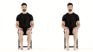 Ankle pumps
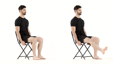 Ankle circles
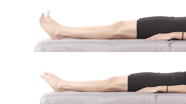 Toe curls
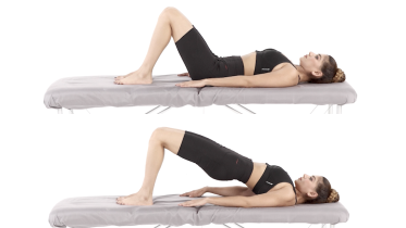 Glute bridge
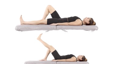 Straight leg raise
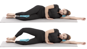 Clamshell
Stage 2: Back in a normal shoe, rebuilding the foot and calf
Start this stage when walking in a normal shoe is pain free and your team is happy for you to load the foot fully. Towel sweeps and scrunches rebuild the arch muscles, which have had weeks off. The two calf raises then rebuild the muscle that absorbs load before it reaches the bones of the foot, starting sitting and progressing to both feet standing. The two calf stretches are for the stiffness that comes from weeks in a boot. Do the seated calf raise with no weight on your knees to begin with, even though the exercise page shows a dumbbell resting there, because anything you put on your knees goes down through the ball of the foot, and add weight only once the bone is no worse the next morning. The standing calf raise is the bigger step of the two, because rising onto the balls of both feet puts your whole body weight through the long bones at the front of the foot, which is where the common cracks are, so start with a small rise and go higher only if the foot stays quiet the next day. Sit to stand is here because standing up from a chair is full weight through both feet, which this stage has cleared, and it keeps the thighs working now that you are off the crutches. Many programs use 2 to 3 sets of 10 to 15, with the strength work 3 days a week and a day between. Your physio will adjust this. Pain the following day means too much, so go back a step rather than pushing on.
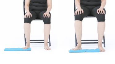 Foot towel sweeps
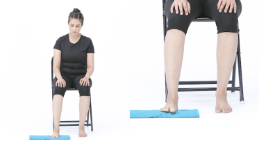 Towel scrunches
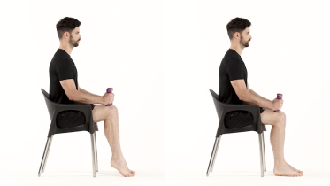 Seated calf raise
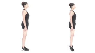 Calf raises
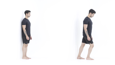 Calf stretch
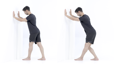 Soleus stretch
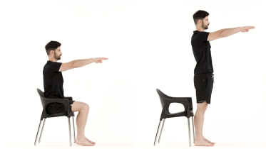 Sit to stand
Stage 3: Single leg strength, walking and balance
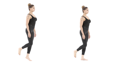
When calf raises on both feet feel easy and walking distances have built back up to what an ordinary day asks of you, without the foot being sore the next morning. If the crack was at the navicular or the base of the fifth bone, do not start this stage on those two criteria alone: standing on one leg sends your whole body weight through both of those bones, so get your surgeon's or fracture clinic's agreement first, the same way stage 4 waits for them. The single leg heel raise is the exercise to measure yourself on: rising onto the injured foot repeatedly shows the calf is taking load again before it reaches the bones of the foot, and it is the one thing in this program you can compare side to side. Its exercise page tells you to push through the big toe side of the foot, which spares the second and third long bones. That is the wrong way round if your crack is in the first long bone behind the big toe or in the small sesamoid bones under it, so for those two keep the weight spread across the ball of the foot instead, without rolling onto the outer edge, and ask your team before you load the foot on one leg at all. Step ups and the squat load the leg as a whole, walking with longer steps undoes the short, careful stride people adopt in a boot, and the balance work restores the steadiness you lose when a foot has been out of action. The longer step drill asks you to land heel first, so if the cracked bone is your heel, keep your usual step length and ask your team before you lengthen it. Keep a counter within reach for the single leg work. Many programs use 2 to 3 sets of 8 to 15 for the single leg heel raise and 2 to 3 sets of 8 to 12 for the step ups and the squat, on 2 to 3 days a week with a day between, with balance holds of 10 to 30 seconds and the walking work most days. Your physio will adjust this. This stage is the biggest jump in load the program asks of the bone, so the stage 2 rule still applies: no pain during it, after it, or the next morning. If the spot that was tender starts to feel tender to press again, stop and go back to your team rather than working through it.
 Single leg heel raise
 Step up
 Squat
 Walking with longer steps
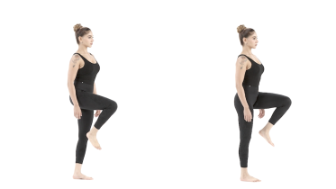 Single leg stance
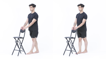 Tandem stance
Stage 4: Impact, and only when the gates are clear
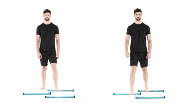
Do not start this stage until the bone is no longer tender to press, you are walking normally and without pain, and your team has cleared you, because this is where bone stress injuries are made worse. If the fracture was at the navicular or the base of the fifth bone, or it was held with a screw or a plate, impact waits for imaging that shows the bone has healed and for your surgeon's word, not for the foot feeling fine. Take them in this order. Pogo hops teach a stiff, quiet landing on two feet. Line jumps stay on two feet but add travel, forward over a line and back again. The single leg hop and hold is the step up from both of those, because it asks one foot to absorb the whole landing, and skater hops come last, since they land on one leg with your body moving sideways. Start with two short sessions a week, well apart, 2 to 3 sets of 5 to 10 landings in each drill to begin with, and build the number of contacts before you build the height or the speed. Your physio will adjust this. Any return of the original pain means stop and go back to your team rather than taking a few days off and trying again. Jumping and hopping also load the joints, the bones and the pelvic floor. If you have osteoporosis or have had a fall in the past year, ask your physio before you do any hopping. If you have had a hip or knee replacement, get the go-ahead from your surgeon before you try hopping. Many surgeons advise against jumping and hopping after a joint replacement for good, not just for the first months, because each landing sends high impact through the new joint. If you are pregnant, have had a baby in the past year, or leak urine when you jump, cough or sneeze, check with your midwife or physio before you start this stage. Stop the hopping and speak to your physio if urine leaks while you are doing it, or if you feel pressure or heaviness low in your pelvis. After having a baby, postnatal guidance usually leaves jumping and hopping until at least 3 months after the birth, once a pelvic floor check shows you are ready. With a heart condition or high blood pressure, check with your doctor before you start.
 Pogo hops
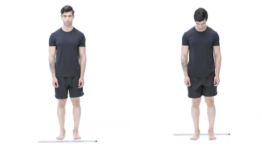 Line jumps
 Single leg hop and hold
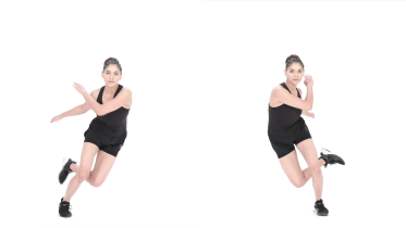 Skater hops
What to train while the foot is offloaded
Six to eight weeks is long enough to lose a noticeable amount of strength and fitness, and almost all of that loss is avoidable. OrthoInfo suggests swimming and cycling as alternatives while the foot heals, with the caveat that nothing involving the injured foot resumes without approval, and the commentary above lists cross-training among the things to use during the offloading period.
In practice it comes down to what you can still train. Keep the hip and thigh working on both legs, which is what stage 1 is for, because a weak hip is one reason people reload the foot badly when they come back. Keep your aerobic fitness with something your team has approved, which is usually cycling with the pedal under the heel, swimming or deep water running. Putting the pedal under the heel is there to spare the long bones at the front of the foot, so it suits a metatarsal injury and is the wrong choice if the crack is in your heel bone, which is the second most common site. And keep training the rest of your body: an upper body session costs the foot nothing.
If you are in a cast or a boot, read the warning signs about tight casts and skin under a cast carefully. Those are the two problems that arise during this phase and neither can be seen from the outside.
Getting back to running and impact
Impact is the point where a healing bone is most easily set back, so the gates matter more than the enthusiasm.
For the decision itself, a scoping review of returning to running after tibial bone stress injuries identified five things to address first: resolution of bony tenderness, pain free walking, radiological healing for the high-risk sites, strength, functional and loading tests, and identifying what contributed to the injury in the first place. Its authors recommend a shared decision between clinician, coach and athlete, and an individual graduated program, often starting with walk-run intervals and progressing distance before speed and intensity (George, Sheerin and Reid, 2024). That review studied the shin rather than the foot, and we are using it for the structure of the decision, not as a foot protocol.
The commentary on low-risk metatarsal and tibial injuries adds the practical trigger: start the return-to-run program once you have been pain free in daily activities for 5 consecutive days, and build volume before speed (Warden, Edwards and Willy, 2021). That trigger belongs to the low-risk sites it was written for, the shin and the shaft of a metatarsal, and not to the navicular or the base of the fifth. OrthoInfo says early rehabilitation may alternate activity days with rest days and that intensity, frequency and duration increase gradually.
Our return to running protocols guide sets out the build-up week by week. If the pain was in the shin rather than the foot, shin splints covers the other end of the same spectrum, and if the pain is spread across the ball of the foot, read metatarsalgia.
Why it happened, and how to make it less likely
OrthoInfo's list of causes is worth reading as a checklist rather than as background: a sudden increase in frequency, duration or intensity, new activities or surfaces, returning to a previous training volume after time off, worn or flimsy shoes, poor conditioning, altered foot mechanics from a blister, a bunion or a tendon problem, and bone insufficiency from osteoporosis or long-term medication. It adds that stress fractures are more common in winter, when vitamin D is lower.
Two of its points deserve more weight than the rest. OrthoInfo says female athletes are more prone than male athletes, possibly due to low bone density related to relative energy deficiency in sport, and the practical meaning is that somebody training hard and eating too little will keep breaking bones until that is addressed. And it advises a bone density scan if the fracture happened with light activity, which is how thin bones get found. If that applies to you, read our osteoporosis page as well.
For prevention it advises calcium and vitamin D in the diet, proper footwear with a softer insole and a stiffer outer sole, replacing worn shoes, gradual increases with a 10% rise per week described as appropriate in most cases, cross-training, and strength training. The 10% figure is a rule of thumb rather than a tested threshold, so use it as a brake rather than a target.
When to see a physio or doctor
Get a foot that is tender over one patch of bone, and worse the more you walk on it, checked rather than managed at home, because the diagnosis changes everything that follows. Get it checked the same day if you cannot put weight on it.
Go back to your team if the pain has not improved over 6 to 8 weeks of doing what they asked, if it returns as soon as you load the foot normally, or if you have had more than one stress fracture. If the tender spot is on the inner side of the top of the midfoot or on the outer edge at the base of the little toe bone, do not wait that long: stop running and jumping now and get those two looked at within a few days.
Check with your doctor before you start exercising if you have a heart or lung condition, or blood pressure that is not under control. If you have diabetes or reduced feeling in your feet, have the foot looked at instead of working through this program alone. Our ankle fracture rehab page covers boots, weight bearing and swelling in more detail if that is where you are.
For physiotherapists
Classify the site as low or high risk before anything else, because the whole plan turns on it, and treat a clear initial radiograph as uninformative rather than reassuring. Both numbers from Warden and colleagues come from low-risk tibial and metatarsal diaphyseal injuries, so neither transfers to the navicular or the proximal fifth, where the immobilization and weight bearing are the surgeon's call. Within those low-risk sites they give a usable load rule, no symptoms during, after or the day following, and a defined entry criterion for return to run at 5 consecutive pain free days in daily activity. George and colleagues give the five-component framework, derived from tibial injuries and level IV evidence throughout, which is worth stating to patients who want certainty.
Screen for low energy availability and menstrual status in every case rather than only in the obvious ones, and recheck it in anyone presenting with a second bone stress injury. Calf endurance, measured as single leg heel raise repetitions compared side to side, is the most useful progress measure this program produces.
Warning signs
Get emergency help now
- Emergency: sudden, severe pain in your lower leg that is much worse than you would expect, especially after an injury or with a plaster cast or tight bandage on the leg. It may come with a tight, swollen leg, pain that gets worse when the muscle is stretched, numbness, pins and needles or weakness. Call emergency services or go to an emergency department straight away, and do not drive yourself. This can be acute compartment syndrome, which needs urgent surgery.
- Emergency: while you are in a cast or boot, your toes turn blue, white or pale, or feel cold. Do not wait to see if raising your leg helps. Go to an emergency department straight away, and do not drive yourself. The cast or boot may be cutting off the blood supply, or pressure may be building up inside the leg (compartment syndrome), and both need checking quickly.
- Part of your foot or a toe turns cold, pale, blue or black. Call emergency services or go to an emergency department straight away, and do not drive yourself, as the blood supply to the foot may be blocked.
Get help now, day or night
- In a cast or boot, the pain gets worse, your toes feel numb, tingle or burn or look more swollen, or the cast or boot feels too tight. Stop, raise your foot to about hip level and no higher, keep moving your toes and contact your surgical team or fracture clinic or get medical advice straight away. If you wear a boot, loosen the straps but keep it on. Do not prop the foot higher than that and do not put ice on it, because if pressure is building up inside the lower leg both of those cut down the blood reaching your foot. If you cannot move your toes, the pain is severe even after painkillers, or the numbness, tingling, burning or swelling has not settled within 30 minutes of raising your foot, go to your fracture clinic or an emergency department straight away, and do not drive yourself.
- Your cast or boot rubs painfully, or there is a blister-like soreness under it, or there is a bad smell, discharge or liquid coming from under the cast, or something has dropped down inside it. Contact your fracture clinic, plaster room or surgical team straight away, and out of hours go to an emergency department rather than waiting for the morning. Skin under a cast can break down where you cannot see it.
Get help today
- Same day: you cannot put weight on the foot, you heard a snap, grinding or popping noise at the time of an injury, or the pain started with a fall or a landing from height. Get it checked the same day.
- Same day: since your operation, your wound is oozing, leaking or has pus coming out of it, or its edges start to come apart, or your foot is tender, swollen, painful or red and it is not getting better or is getting worse, or you have a high temperature or feel hot and cold or shivery. Redness can be harder to see on brown or black skin. Contact your surgical team the same day, or get urgent medical advice if you cannot reach them. These can be signs of an infection in the wound, or deeper in a joint, a bone or around metalwork in the foot. If you feel very unwell, go to an emergency department. If you have a very high or very low temperature, shivering you cannot control, you are breathing very fast or finding it hard to breathe, you are confused or your speech is slurred, your skin, lips or tongue look blue, gray, pale or blotchy (on brown or black skin this can be easier to see on the palms of the hands or soles of the feet), or you have a rash that does not fade when you press it, call emergency services, as this can be sepsis.
- Same day: since your operation, you felt a crack, snap or sudden give in the operated foot, or it has started to give way under you, or you suddenly cannot put weight through it when you could before, or the shape of the foot is changing, with the arch dropping again, a toe that was straightened shifting back, or the foot starting to look out of line or to sit differently in a boot or cast, or a boot or cast that fitted starts to feel loose and roomy. Bones that were cut, moved or fused, and tendons that were repaired or moved, are held in place by screws, plates, wires or stitches while they knit, and a correction can slip without any new injury and without a snap. It is put right more easily when it is found early. Stop the exercises, keep your weight off that foot and contact your surgical team the same day, or get urgent medical advice if you cannot reach them. If the foot looks clearly out of shape, or you cannot stand on it and cannot reach anyone, go to an urgent care center or an emergency department.
- The ankle or foot is hot, red and swollen, or you have a fever or feel unwell. Get medical help the same day, and go to an emergency department if you feel very unwell. If you have a very high or very low temperature, shivering you cannot control, you are breathing very fast or finding it hard to breathe, you are confused or your speech is slurred, your skin, lips or tongue look blue, gray, pale or blotchy (on brown or black skin this can be easier to see on the palms of the hands or soles of the feet), or you have a rash that does not fade when you press it, call emergency services, as this can be sepsis.
- Same day: you have diabetes or numb feet, and one foot becomes hot, swollen or red, or changes color or shape, with or without pain. Take your weight off that foot straight away and get advice the same day from your doctor or your diabetes foot team. This can be Charcot foot, where the bones and joints of the foot are damaged and the foot can collapse. Do not exercise that foot until it has been checked. If you also have a fever or feel very unwell, or redness is spreading up your foot or leg, go to an emergency department.
- Same day: you have diabetes or numb feet and find a new blister, cut or sore on your foot, especially one you did not feel, a wound that smells or is leaking, or new pain in your foot that you cannot explain. Get it seen by your doctor or foot team the same day. If you also have a fever or feel very unwell, redness is spreading up your foot or leg, or part of your foot or a toe turns black, go to an emergency department.
- A calf or thigh that is newly swollen or getting more swollen, warm, tender, red or darker than usual, has swollen veins that are sore to touch, or has a throbbing or cramping pain that does not fit your injury or condition, or that feels different from normal muscle soreness after exercise. This can be a sign of a blood clot (deep vein thrombosis), especially after surgery, an injury, time in a cast or boot, a long trip or a spell of being much less mobile than usual. Get medical advice the same day. If you are also short of breath or have chest pain, call emergency services.
- Same day: your cast feels too loose or uncomfortable, cracks, softens, gets wet or itches badly. Ring your surgical team, fracture clinic or plaster room and ask to be seen, and go to an emergency department if they cannot see you that day. A cast that has gone loose or soft may no longer be holding the broken bone still, so keep your weight off that leg until you have been seen, and do not cut, pad or adjust it yourself.
Book an appointment
- Since the fracture or cast, your foot or ankle is getting more painful rather than less, often with a burning pain that feels far worse than the injury, the skin becomes very sensitive to light touch, bedclothes or a change in temperature, or the foot changes color, temperature, swelling or sweating compared with the other one, or looks shiny. See your doctor or physio as soon as you can, within a few days at most, as this can be complex regional pain syndrome (CRPS) and early treatment may help.
- Your pain sits over the bony bump on the inner side of the top of the midfoot, or on the outer edge of the foot at the base of the little toe bone, and it is deep, hard to pinpoint, or worse with sprinting and jumping. Stop running and jumping now, keep your weight off the foot as much as you can, and see a doctor within a few days. Say which spot it is when you book, and get it checked the same day instead if walking hurts or makes you limp. The navicular in the midfoot and the base of the fifth bone are the two sites that heal slowly and unpredictably, and they are the two most likely to fail to knit and end up being held with a screw, so they are confirmed and managed properly rather than waited out. OrthoInfo says the base of the fifth has a poor blood supply, that treatment of a navicular stress fracture will require a period of taking no weight at all through the foot, and that an early X-ray is often normal because the crack is too fine to show, so a clear film is not the end of it. Ask what imaging settles it, which for the navicular is a CT or an MRI scan.
- Your foot pain has not improved after 6 to 8 weeks of the offloading and footwear your team advised, or it comes straight back as soon as you walk or run normally again. This is not an emergency, but go back to the offloading your team set rather than carrying on walking or running on it, and ask your doctor or fracture clinic for a review in the next week or two, or within a few days if the painful spot is the navicular or the base of the fifth bone. Ask whether the foot needs imaging again. Whether a bone has united is decided on a scan and not on how the foot feels, so a foot that has gone quiet is not proof, and a stress fracture that is not uniting needs a different plan rather than more time. Keep every follow-up appointment, including the scans, and if you have already been discharged and the same spot is still tender, ask to be sent back instead of waiting to see whether it settles.
- You have had more than one stress fracture, or this one came on with light activity rather than hard training, or you have been eating less than your training needs or losing weight, or you are not having periods or your periods have stopped since you started training harder. Ask your doctor for a review in the next week or two and mention all of it together rather than one piece at a time. OrthoInfo advises considering a bone density scan when a stress fracture happens with light activity, and blood tests for calcium and vitamin D if you have had a stress fracture before, and low energy availability in sport is treatable once somebody is looking for it. Raise it even if it feels like an awkward thing to bring up, because this is the part that decides whether you get the next one.
Common questions
How long does a foot stress fracture take to heal?
OrthoInfo says healing typically takes 6 to 8 weeks, and that more serious fractures, chronic ones, and fractures in bone with less blood supply take longer. The exception it names specifically is the base of the fifth metatarsal on the outer edge of the foot, where it advises non-weightbearing immobilization in a cast or boot for at least 6 weeks and says return to sports is usually not until 12 weeks after the injury. For the navicular it gives no timeline at all, and a systematic review of published series found an average of about 22 weeks back to sport when it was treated without surgery and about 16 weeks when it was fixed, on very low quality evidence (Mallee and colleagues, 2015). So 6 to 8 weeks is the figure for the common sites, and it does not cover the two awkward ones. Ask your team which bone it is before you plan anything.
What are the symptoms of a foot stress fracture?
OrthoInfo describes pain that develops gradually and gets worse with weight bearing, that goes away during rest or comes with activities through the day, swelling on the top of the foot or outside of the ankle with possible bruising, and tenderness when pressure is put directly over the bone. That last one is the useful sign: a stress fracture is usually tender over a small patch of bone you can find with one finger, rather than achy across a whole area. The site changes the picture, and OrthoInfo notes that heel stress fractures can feel like plantar fasciitis, navicular pain is hard to pinpoint and worse with sprinting or jumping, and sesamoid pain is felt around the base of the big toe.
How is a foot stress fracture diagnosed?
By examination and then imaging, and the important thing to know is that a normal X-ray does not rule it out. OrthoInfo says X-rays can be normal early and the fracture may not be visible until several weeks later, when a callus appears. An MRI or a bone scan is more sensitive and can detect it earlier, with MRI more specific for the location, and a CT scan is used for surgical planning or to check whether a stress fracture has become a complete break. If your X-ray was clear and the bone is still tender in one spot, that is a reason to go back, not a reason to train on.
Can you walk on a foot stress fracture?
Follow your team's instruction, because the answer depends on the bone. OrthoInfo describes crutches to keep weight off the foot until the pain settles, then footwear that stops the foot bending: a shoe with a rigid sole, a sandal built on a wooden base, or a removable brace that takes in the lower leg as well as the foot. Fractures that need steadier holding may be put in a cast instead. Two sites are different: for the base of the fifth metatarsal OrthoInfo advises non-weightbearing immobilization for at least 6 weeks, and it says treatment of a navicular stress fracture will require a period of non-weightbearing too, so for either of those the answer is no until your team says otherwise. Walking on a stress fracture that hurts is how it becomes a complete break, which is also why the warning signs on this page include being unable to put weight on the foot.
What exercise can I do with a foot stress fracture?
Most things that do not load the foot. OrthoInfo suggests swimming and cycling while the foot is healing, and adds that no activity involving the injured foot should resume without approval. Stage 1 of the program above keeps the hip and thigh working on both legs, which is the part people skip and then regret, because six weeks of doing nothing costs you strength everywhere. Upper body work, a stationary bike with the pedal under the heel if your team allows it, and deep water running are the usual choices. The pedal under the heel is there to keep load off the long bones at the front of the foot, so it is the wrong way round if the cracked bone is your heel. Ask before you start any of them, because what is safe depends on the bone.
When can I start running again after a foot stress fracture?
On how the foot behaves, not on a date. A clinical commentary in the Journal of Orthopaedic and Sports Physical Therapy on low-risk tibial and metatarsal bone stress injuries in runners suggests starting a return-to-run program once the runner has been pain free during daily activities for 5 consecutive days, and progressing volume before speed (Warden, Edwards and Willy, 2021). A scoping review of return to running after tibial bone stress injuries lists five things to settle first: no bony tenderness, pain free walking, radiological healing for the high-risk sites, strength and loading tests, and identifying what caused it (George, Sheerin and Reid, 2024). That review is about the shin rather than the foot, so take it as the shape of the decision rather than a foot-specific protocol. Neither piece of work covers the navicular or the base of the fifth metatarsal: the commentary is about low-risk sites, OrthoInfo puts return to sport after a fifth metatarsal base fracture at usually not before 12 weeks, and if yours is one of those two the date is your surgeon's or your fracture clinic's and not this page's.
Why did I get a foot stress fracture?
Usually because the loading went up faster than the bone could adapt to it. OrthoInfo names a sudden increase in activity, including returning to a previous training volume after time off, a change of surface such as grass to hard court or treadmill to track, worn-out or flimsy shoes that have lost their shock absorption, poor conditioning, and altered foot mechanics from a blister, a bunion or a tendon problem. It also names bone insufficiency from osteoporosis or long-term medications, says stress fractures are more common in winter when vitamin D is lower, and says female athletes are more prone than male athletes, possibly through low bone density related to relative energy deficiency in sport. Non-athletes get them too, and OrthoInfo gives the example of somebody walking much more than usual on vacation.
Does a foot stress fracture need surgery?
Most do not. OrthoInfo says some need stabilizing, usually with internal fixation using pins, screws or plates, and names the two situations: the awkward sites, where it mentions a screw, bone grafting or plates and screws for the base of the fifth metatarsal and says surgery is more commonly recommended for the navicular, and a stress fracture that has broken completely with the pieces displaced. It adds that the hardware usually does not need removing. If an operation has been suggested, the weight bearing and return to impact dates come from your surgeon and this page does not set them.
How do I stop it happening again?
Fix the loading, the fuel and the shoes, in that order. OrthoInfo advises a diet rich in calcium and vitamin D, proper footwear with a softer insole and a stiffer outer sole with worn shoes replaced, cross-training so one area is not overstressed, adding strength training, and increasing activity gradually, saying that a 10% increase per week is appropriate in most cases. Treat that percentage as a rule of thumb rather than a tested threshold. The two things most worth your attention are keeping the calf strong, because it absorbs load before the foot bones do, and eating enough for the training you are doing, because a stress fracture in somebody training hard and eating too little will keep coming back whatever the shoes are like.
References
- American Academy of Orthopaedic Surgeons. Stress Fractures of the Foot and Ankle. OrthoInfo. https://www.orthoinfo.org/en/diseases--conditions/stress-fractures-of-the-foot-and-ankle/
- Warden SJ, Edwards WB, Willy RW. Optimal Load for Managing Low-Risk Tibial and Metatarsal Bone Stress Injuries in Runners: The Science Behind the Clinical Reasoning. Journal of Orthopaedic and Sports Physical Therapy. 2021;51(7):322-330. https://doi.org/10.2519/jospt.2021.9982
- George ERM, Sheerin KR, Reid D. Criteria and Guidelines for Returning to Running Following a Tibial Bone Stress Injury: A Scoping Review. Sports Medicine. 2024;54(9):2247-2265. https://doi.org/10.1007/s40279-024-02051-y
- Mallee WH, Weel H, van Dijk CN, van Tulder MW, Kerkhoffs GM, Lin CW. Surgical versus conservative treatment for high-risk stress fractures of the lower leg (anterior tibial cortex, navicular and fifth metatarsal base): a systematic review. British Journal of Sports Medicine. 2015;49(6):370-376. https://doi.org/10.1136/bjsports-2013-093246
- Troy KL, Davis IS, Tenforde AS. A Narrative Review of Metatarsal Bone Stress Injury in Athletic Populations: Etiology, Biomechanics, and Management. PM&R. 2021;13(11):1281-1290. https://doi.org/10.1002/pmrj.12518
- NHS. Compartment syndrome. Page last reviewed 4 September 2026. https://www.nhs.uk/conditions/compartment-syndrome/
- NHS. DVT (deep vein thrombosis). Page last reviewed 30 April 2026. https://www.nhs.uk/conditions/deep-vein-thrombosis-dvt/
- North Bristol NHS Trust. Care of plaster casts. Patient information, October 2024. https://www.nbt.nhs.uk/care-of-plaster-casts
Written and checked by the PocketPhysio editorial team. Last updated 2026-10-28.
This page is general education, not a diagnosis or a personal treatment plan. See a physiotherapist or doctor for advice about your own situation.