
Dislocated kneecap (patellar dislocation) exercises and physiotherapy
What happens when a kneecap dislocates
Your kneecap (patella) glides up and down in a groove at the end of the thigh bone as you bend and straighten the knee. In a dislocation it comes right out of that groove (Smith and colleagues, 2023), and the research on this page is about dislocations toward the outer side of the knee (lateral dislocations). The NHS says it is usually caused by an injury, and that it often goes back into place by itself. A dislocation can stretch or tear the band on the inner side that holds the kneecap in its groove, the medial patellofemoral ligament (MPFL). It can also break off a piece of cartilage or bone inside the joint, which happens often, especially in children and teenagers (Blønd and colleagues, 2025).
So the knee needs checking even if the kneecap went straight back in. A European consensus of knee experts (ESSKA) considers an X-ray and an MRI scan, or an MRI alone, necessary soon after a first dislocation in most cases, to look for broken pieces and for the knee shapes that make another dislocation more likely (Blønd and colleagues, 2025). A knee that fills with blood soon after the injury is a warning sign for a broken piece.
This page is for adults after a first dislocation that has been checked and is being treated without surgery. A child or teenager who is still growing needs their own assessment by a doctor or physio, so do not use this program for them. If you are pregnant, check with your midwife, doctor or physio before you start. If you have had surgery on your kneecap, follow the program from your surgeon and physio instead.
What to do straight after a dislocation
If the kneecap is still out of place, do not try to push it back in yourself. The NHS advice is to keep the knee still, support it with something soft, not stand or walk on it, and go to an emergency department, or call emergency services if you cannot move the injured leg. Do not drive yourself.
At the hospital, a doctor gives strong pain relief before moving the kneecap back into place if it has not gone back by itself, and you usually have X-rays (NHS). You may be given a crutch if walking is painful, and a knee support. For the first days at home, the NHS suggests ice packs for 10 to 15 minutes at a time, at least 3 times a day, painkillers such as paracetamol or ibuprofen, and resting with the leg raised and supported.
Is this the same as patellofemoral pain?
No, although the two can overlap. Patellofemoral pain is an ache around or behind the kneecap that builds up gradually, with no injury and without the kneecap coming out. This page is for a knee where the kneecap has actually come out of place, so the program adds early protection, single leg control and a careful return to landing and turning. Some people are left with kneecap pain after a dislocation. If your knee has become stable and the main problem is an ache on stairs, squats or long spells of sitting, ask your physio whether the patellofemoral pain program suits you better.
Does physiotherapy help after a dislocated kneecap?
It is the usual treatment, but honestly, the evidence behind the exercises themselves is thin. The ESSKA consensus recommends physio-guided exercise alongside both surgical and non-surgical treatment, while stating that strong scientific evidence for it is lacking (Balcarek and colleagues, 2025). It lists what that exercise should cover: strengthening the thigh and buttock muscles (quadriceps and gluteal muscles), getting the knee moving, walking normally again, control of the leg in functional movements, and sport-specific training where needed. The stages below follow that outline, and your physio will fit them to your knee and your sport.
Keep in mind who wrote it. The ESSKA group was 54 orthopedic surgeons and 1 physiotherapist from 20 European countries (Blønd and colleagues, 2025), and none of its statements on treatment reached the top grade of evidence. Much of it is expert agreement where trials are missing.
Bracing, crutches and the first weeks
The NHS says you may need a knee support and a crutch at first. The ESSKA consensus found no evidence that any brace does better than no brace, early or later, and says a brace that lets the knee bend freely may be considered only for a very short time after the injury (Balcarek and colleagues, 2025). So wear what your doctor gave you, as they advised, and expect to use it for a short time.
Walking is usually allowed early. The NHS notes that many people can start walking again as soon as they have been treated, and advises keeping active. Keep the knee moving gently within the range that feels comfortable, and let the swelling guide you.
How to use this program
Start at the stage that fits how your knee is now. Not sure? Begin with stage 1. Move on when the stage you are on feels easy and the knee is no worse the morning after. Treat these signs as a rough guide from common practice rather than fixed rules.
You will find a typical starting dose on each exercise page. As a rough guide, many programs use 2 to 3 sets of 6 to 15 repetitions for the strength exercises, with the lower numbers for the harder single leg work, once a day or a few days a week. Quad sets and heel slides are often done little and often, 2 or 3 times a day or more. Wall sit holds usually start at 10 to 30 seconds, repeated 3 to 5 times. If you have high blood pressure, breathe steadily through every hold and never hold your breath.
Jumps and hops usually start with short sets, often 5 to 10, with a full rest between them, a few times a week. Your physio will set the exercises and the numbers for your knee.
In stage 1, stay pain free. From stage 2 on, some mild discomfort during the exercises is usually fine, as long as it settles soon after you finish and the knee is no worse the next morning. Sharp pain, more swelling the next day, or any feeling that the kneecap is shifting or about to slip means the load was too much. Ease off rather than stopping altogether: a shallower squat or a lower step is often enough.
The exercise program
Stage 1: Protect the knee and get it moving
For the first days and weeks after the kneecap has gone back in and been checked, while the knee is swollen and you may be in a support or on crutches. Put as much weight on the leg as feels comfortable, unless your doctor has told you otherwise, and use the crutches while walking still makes you limp. These exercises are done lying or sitting, so the front of the thigh (quadriceps) and the side of the hip start working again without any twisting through the knee. Slide the heel only as far as is comfortable, and keep this stage pain free.
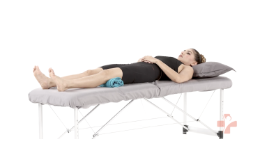 Quad sets (towel under knee)
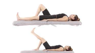 Straight leg raise
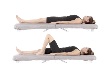 Heel slides
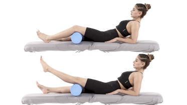 Short arc quads
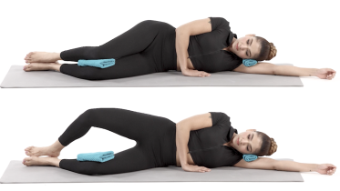 Clamshell
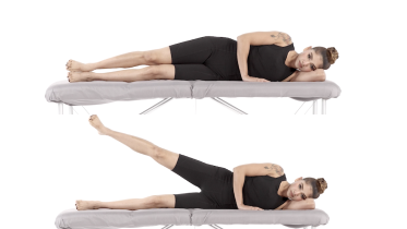 Side lying hip abduction
Stage 2: Build strength with weight on the leg
When you can walk without crutches and without a limp, the swelling has mostly settled, you can lift your leg straight without the knee sagging, and stage 1 feels easy. The seated and band exercises build the front of the thigh, and the bridge, mini squat, wall sit and calf raises train the leg with your weight on it. Start the squat and the wall sit high and shallow, go only as deep as feels steady, and keep your kneecap pointing over your second toe so the knee does not drift inward. Do the single leg stance next to a kitchen counter you can hold.
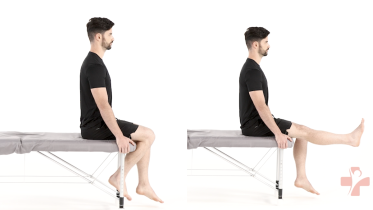 Seated knee extension
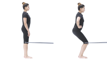 Terminal knee extension with band
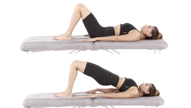 Glute bridge
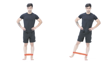 Standing hip abduction with band
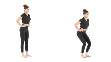 Mini squat
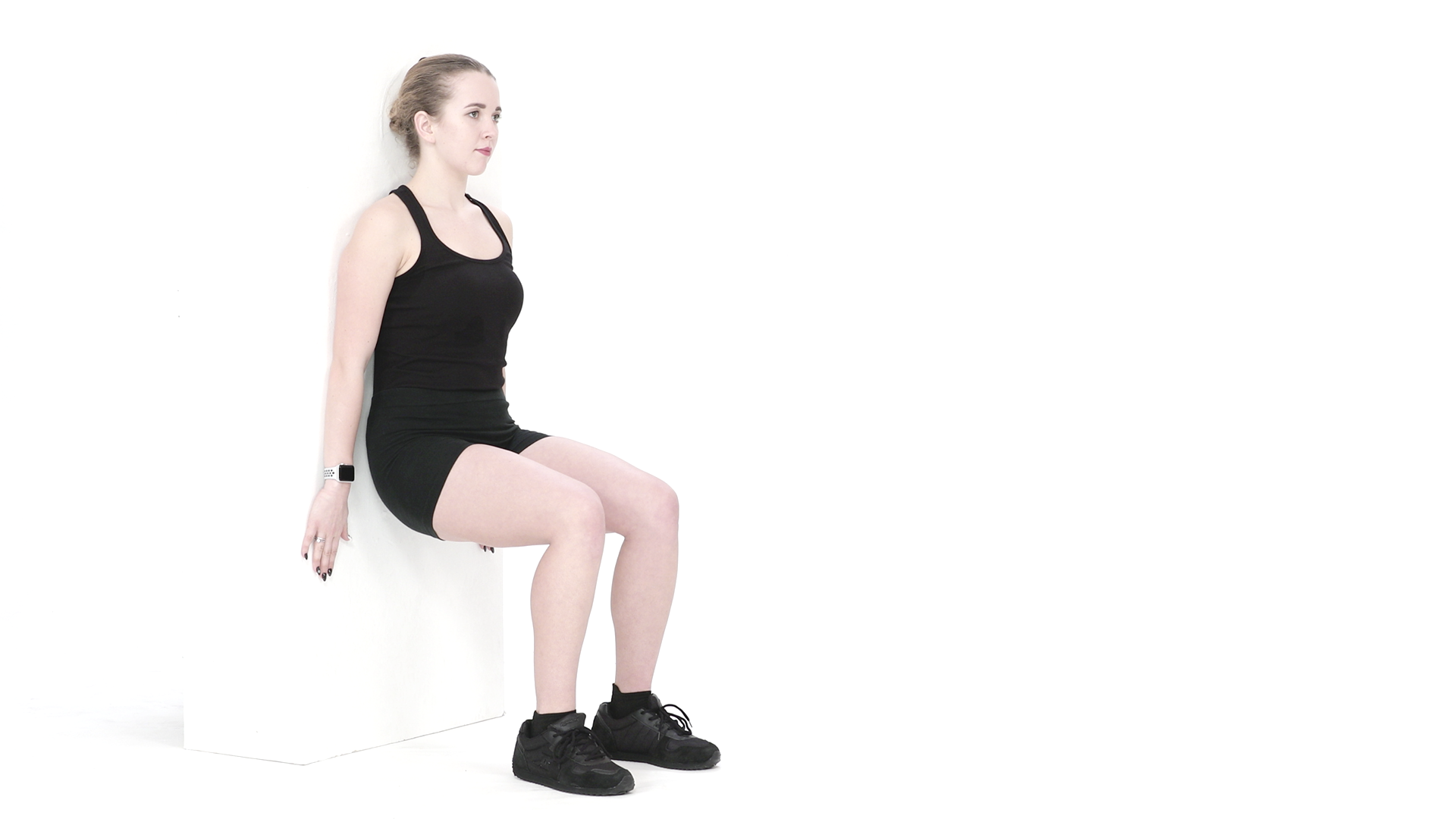 Wall sit
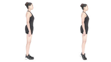 Calf raises
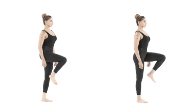 Single leg stance
Stage 3: Single leg strength and control
When stage 2 feels easy, the swelling has gone and the knee is no worse the next morning. These exercises ask one leg to hold the knee in line while you step, lunge and balance, which is what stairs, running and sport need. Use a low step for the step up and the step down, and start the split squat and lunge with a short range. The band walk and the single leg bridge work the side and back of the hip, and the single leg deadlift adds balance as you hinge forward. Keep a counter or wall within reach for all the single leg work.
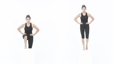 Step up
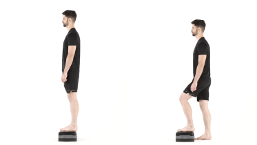 Backward step-down
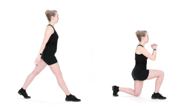 Split squat
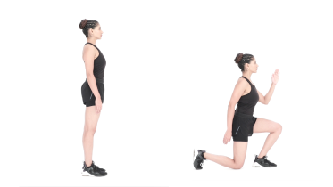 Reverse lunge
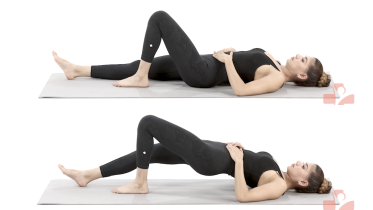 Single leg bridge
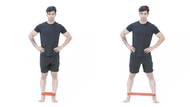 Lateral band walk
.png) Single leg deadlift
Single leg deadliftStage 4: Jumping, hopping and the road back to sport
When single leg work feels solid, the knee bends and straightens fully and no longer swells after exercise, and the kneecap no longer feels as if it might slip. Start with jumps on two feet, then hops on one, and only then the side lunge and skater hops, which move you sideways and land you on one leg. If you have osteoporosis or have had a fall in the past year, ask your physio before you do any jumping or hopping. For sports with cutting and turning, your physio can add running drills with changes of direction and plan the last steps back to full training.
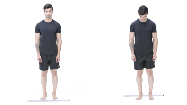 Line jumps
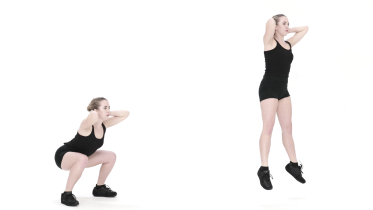 Jump squats
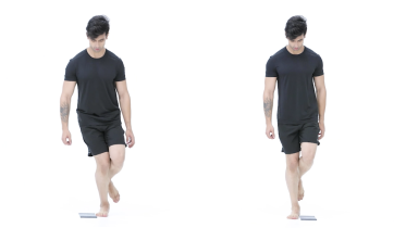 Single leg hops over a line
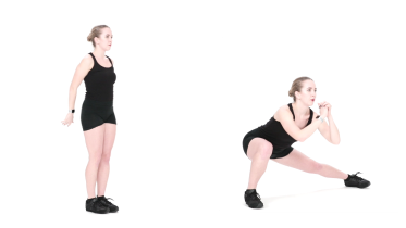 Lateral lunge
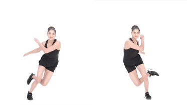 Skater hops
What to avoid while the knee recovers
The NHS advises avoiding twisting the knee while it recovers. Many physios also hold back pivoting on a planted foot until the thigh and hip are stronger. Deep squats and kneeling wait too, as does any movement where the knee drifts inward past the foot. This comes from common practice rather than from trials. Stages 3 and 4 bring back deeper bending and sideways work, and then landing, once the knee copes with straight line work.
Will my kneecap dislocate again?
It might, and for some knees the risk is high. A review of 17 studies found that about a third of people (33.6%) had another dislocation after a first one (Huntington and colleagues, 2020). Being younger, still growing, having a shallow groove for the kneecap (trochlear dysplasia), a kneecap that sits high (patella alta) or a shin bone bump that sits further out (a raised TT-TG distance) all raised the risk. Being male or female made no clear difference.
The risk factors add up. With none of them, 7.7% to 13.8% dislocated again, rising to 29.6% to 60.2% with 2 risk factors and to 70.4% to 78.5% with 3 (Huntington and colleagues, 2020). The ESSKA consensus says at least 1 in 4 people treated without surgery dislocate again (Balcarek and colleagues, 2025), and that about half of people have other lasting problems such as pain, swelling, giving way or playing less sport (Blønd and colleagues, 2025). Your X-ray and scan show which risk factors apply to you, so ask your doctor to explain them.
When is surgery (MPFL reconstruction) considered?
After a first dislocation in an adult, the ESSKA consensus supports treatment without surgery when the risk of it happening again is low and no cartilage or bone has broken off (Balcarek and colleagues, 2025). It suggests surgery as a first option when a person has several risk factors and ongoing symptoms, or a broken piece of cartilage or bone. The NHS lists a badly damaged knee, or a kneecap that keeps dislocating, as reasons for an operation.
The operation the consensus prefers rebuilds the ligament on the inner side of the kneecap, which is called MPFL reconstruction. It rates it above stitching the torn ligament and says the surgeon may also correct the shape of the bones at the same time when that is part of the problem (Balcarek and colleagues, 2025). A larger broken piece of cartilage or bone, about 1 square centimeter or more in the part of the joint that takes load, should be repaired rather than removed.
A Cochrane review of 10 trials with 519 people shows how unsure the evidence still is (Smith and colleagues, 2023). If about 35 in 100 people treated without surgery dislocated again within 2 to 9 years, about 16 fewer in 100 did so after surgery, but the certainty was very low. Satisfaction was much the same in both groups, the review could not say whether surgery improved knee function or caused more complications, and no large enough trial has looked at people whose kneecap has dislocated more than once. The decision is one to make with a knee surgeon, and if you do have surgery, follow the program your surgeon gives you rather than this page.
Getting back to sport
Most people want a date, and there is no fixed one. The NHS advises no sport until your knee has fully recovered its usual strength, and recovery can take several months. Many physios want full movement and no swelling first, with strength and hopping close to the other leg. You should also be able to run and turn with confidence, without the kneecap feeling as if it will slip. That is common practice rather than a tested set of rules, and your physio will plan the last steps with you.
When to see a physio or doctor
Any first kneecap dislocation needs checking by a doctor, even if it went back by itself, so a broken piece of cartilage or bone is not missed. After that, a physical therapist (physiotherapist) can guide the program, and it is worth seeing one if you were not referred. See your physio or doctor again if the kneecap feels as if it will slip during everyday tasks or sport, slips out partly and goes back, or dislocates again, as that can change the advice about surgery.
It is also worth a check if the knee is not improving as you expected after a few months, still swells with activity, or you are not sure which stage suits you. Some problems need help sooner, anything from an emergency visit to an appointment within a few days, and the warning signs below say which. Check with your doctor before you start if you have a heart or lung condition, or blood pressure that is not under control.
For physiotherapists
This page gives patients a starting framework for nonoperative management after a first-time lateral patellar dislocation in a skeletally mature patient. It does not cover recurrent instability or postoperative rehab.
ESSKA 2024 Part 1 (Blønd and colleagues, 2025) recommends prompt radiographs (AP, lateral and axial) and MRI, or MRI alone, to rule out osteochondral fractures and assess trochlear dysplasia, patella alta and axial alignment, and treats hemarthrosis or lipohemarthrosis as a warning sign. It also notes an inverse relationship between trauma intensity and underlying pathoanatomy. It asks for the mechanism to be recorded along with age and family history, and whether there are bilateral instability symptoms. Once the acute effusion settles, it suggests testing patellar glide and apprehension and looking for a J-sign.
In Part 2 (Balcarek and colleagues, 2025), nonoperative care is supported in skeletally mature patients with low recurrence risk and no chondral or osteochondral lesion (grade C). There is no evidence for any brace over no brace, with a short period of unrestricted range of motion bracing at most in the acute phase (grade B). Physio-guided exercise covering quadriceps and gluteal strengthening, range of motion, gait re-education, functional neuromuscular control and sport-specific training is recommended despite weak evidence (grade C). Primary surgical stabilization is suggested as a first option with several risk factors and ongoing symptoms, or with osteochondral lesions (grade C), with MPFL reconstruction preferred over repair (grade C) and chondral or osteochondral defects of 1 cm2 or more in the contact area repaired (grade C).
Huntington and colleagues (2020) pooled 17 studies and reported odds ratios for recurrence of 2.61 for younger age, 2.72 for open physes, 4.15 for trochlear dysplasia, 2.87 for raised TT-TG distance and 2.38 for patella alta, with no association for sex, MPFL injury pattern or a history of contralateral dislocation. The 2023 Cochrane update (Smith and colleagues) rated every outcome very low certainty, with a Kujala difference of 5.73 points favoring surgery (95% CI 2.91 lower to 14.37 higher) against a 10-point threshold for a meaningful change. Progression here depends on the 24-hour response to load and on effusion and apprehension, not on fixed timelines. Valgus and rotational loading come last.
See a doctor promptly if
- Emergency: the kneecap is still out of place, the knee has changed shape or looks misshapen, or there is heavy bleeding or bone showing through the skin. Go to an emergency department straight away, and do not try to push the kneecap back in yourself. Do not drive yourself: ask someone to drive you, or call emergency services if you cannot get there or cannot move the injured leg.
- Emergency: after a knee injury, you have tingling or no feeling in the leg, foot or toes, or your foot turns cold, pale or blue. Call emergency services or go to an emergency department straight away, and do not drive yourself. These are not usual after a kneecap dislocation, and the blood supply to the leg or a nerve may have been damaged.
- Emergency: after a new injury, the knee swelled up very quickly or you felt a pop or snap. Go to an emergency department straight away. Fast swelling usually means bleeding inside the joint, and after a kneecap dislocation it can mean a piece of cartilage or bone has broken off, which needs an X-ray and usually a scan.
- Same day: the kneecap slipped out of place and went back by itself, whether it is the first time or it has happened before. Get medical advice the same day, even if the knee feels better, so it can be checked for a broken piece of cartilage or bone.
- Same day: the knee is locked, which means it is stuck and you cannot fully straighten it, even after gently trying to move it, or it keeps catching. Get assessed the same day by a doctor, at urgent care or at an emergency department. A piece of cartilage or bone that broke off during the dislocation can get caught in the joint and block it.
- Same day: the knee swells up quickly and a lot with no clear reason, especially if you take medicine to thin your blood. Get medical advice the same day.
- Same day: you cannot walk or put weight on the leg, you cannot stand up or move the knee, or the pain is very bad after a fall or injury. Get medical advice the same day. If you cannot move the injured leg at all because of the pain, call emergency services.
- Same day: the knee keeps giving way under you, or it gave way after a recent injury, or it gives way and is also very painful or swollen. Get medical advice the same day. If it only happens now and then, with no recent injury and no swelling, it is not an emergency, but book an assessment with a physio or doctor in the next few days.
- Same day: the knee is hot, red and swollen, or you have knee pain with a high temperature or feel hot, cold or shivery. Get medical advice the same day, and go to an emergency department if you feel very unwell. This can be a sign of infection in the joint, which needs treatment quickly.
- Same day: a calf or thigh that is newly swollen, warm, tender, red or darker than usual, has swollen veins that are sore to touch, or has a throbbing or cramping pain that does not fit your injury or condition, or that feels different from normal muscle soreness after exercise. This can be a sign of a blood clot (deep vein thrombosis), especially after surgery, an injury, time in a cast or boot, a long trip or a spell of being much less mobile than usual. Get medical advice the same day. If you are also short of breath or have chest pain, call emergency services.
- Within a few days: pain at night or at rest that keeps getting worse. See your doctor within a few days. If you have had cancer, now or in the past, see your doctor sooner, within a day or two, and mention it, even if the pain is not getting worse. Do the same if you have knee pain and are losing weight without trying, even if it does not hurt at night. If you are being treated for cancer now, contact your cancer team the same day.
Common questions
How long does a dislocated kneecap take to heal?
The NHS says it can take several months to fully recover, although many people can start walking again as soon as they have been treated. Getting back to sport usually takes the longest, because strength, hopping and turning have to come back first, and the NHS advises no sport until the knee has its usual strength again. Your physio will judge readiness from how your knee performs, not from the calendar alone.
Will my kneecap dislocate again?
It might, and the risk depends a lot on the shape of your knee and your age. In a review of 17 studies, about a third of people had another dislocation after a first one (Huntington and colleagues, 2020). With none of the main risk factors, the rate was about 8 to 14 in 100, and with 3 of them it was over 70 in 100. Your doctor can explain your own risk from your X-ray and scan.
Do I need surgery for a dislocated kneecap?
Not always. After a first dislocation, a European consensus of knee experts supports treatment without surgery when the risk of another dislocation is low and no piece of cartilage or bone has broken off. The same consensus names MPFL reconstruction as the operation of choice when surgery is needed (Balcarek and colleagues, 2025). A Cochrane review found surgery may lower the chance of another dislocation, but the evidence was very low certainty and it could not say whether surgery gives a better result overall (Smith and colleagues, 2023). A knee surgeon makes that call with you.
Should I wear a knee brace after a dislocated kneecap?
Only for a short time, if at all. The same European consensus found no evidence that any brace works better than no brace, and says a brace that lets the knee bend freely may be used for a very short time just after the injury (Balcarek and colleagues, 2025). The NHS notes you may be given a knee support. Wear it as your doctor or physio advised, and do not rely on it in place of the exercises.
Can you walk on a dislocated kneecap?
Not while it is still out of place: keep the knee still and go to an emergency department. Once it is back in and has been checked, the NHS says many people can start walking straight away, with a crutch if walking hurts. Put as much weight on the leg as feels comfortable, and let your physio know if the knee gives way.
References
- NHS. Dislocated kneecap. Page last reviewed 10 March 2026. https://www.nhs.uk/conditions/dislocated-kneecap/
- Smith TO, Gaukroger A, Metcalfe A, Hing CB. Surgical versus non-surgical interventions for treating patellar dislocation. Cochrane Database of Systematic Reviews. 2023;1(1):CD008106. https://doi.org/10.1002/14651858.CD008106.pub4
- Huntington LS, Webster KE, Devitt BM, Scanlon JP, Feller JA. Factors associated with an increased risk of recurrence after a first-time patellar dislocation: a systematic review and meta-analysis. American Journal of Sports Medicine. 2020;48(10):2552-2562. https://doi.org/10.1177/0363546519888467
- Blønd L, Askenberger M, Stephen J, Akmeşe R, Balcarek P, et al. Management of first-time patellar dislocation: the ESSKA 2024 formal consensus, Part 1. Knee Surgery, Sports Traumatology, Arthroscopy. 2025;33(5):1925-1932. https://doi.org/10.1002/ksa.12620
- Balcarek P, Blønd L, Beaufils P, Askenberger M, Stephen JM, et al. Management of first-time patellar dislocation: the ESSKA 2024 formal consensus, Part 2. Knee Surgery, Sports Traumatology, Arthroscopy. 2025;33(12):4197-4206. https://doi.org/10.1002/ksa.12637
- NHS. Knee pain. Page last reviewed 21 December 2023. https://www.nhs.uk/symptoms/knee-pain/
- NHS. Deep vein thrombosis (DVT). Page last reviewed 30 April 2026. https://www.nhs.uk/conditions/deep-vein-thrombosis-dvt/
Written and checked by the PocketPhysio editorial team. Last updated 2026-09-28.
This page is general education, not a diagnosis or a personal treatment plan. See a physiotherapist or doctor for advice about your own situation.