
Plica syndrome knee exercises and physiotherapy
Warning signs, and when to get help
What is a plica, and what is plica syndrome of the knee?
A plica is a fold in the lining of the knee joint, left over from how the knee forms before birth. Most knees have at least one. The one that causes trouble is usually the medial patellar plica, a shelf of tissue running down the inner side of the kneecap, and Kuwabara and Fredericson note that medial plicae are both the most common and the most often symptomatic (Kuwabara 2021).
Plica syndrome is the name for pain coming from one of those folds when it becomes thickened or irritated and starts catching between the kneecap and the thigh bone. The pattern described in the literature is pain localized to the fold with snapping, clicking, catching, clunking, grinding or popping inside the knee as it bends and straightens, brought on by repeated bending and straightening or by long spells with the knee held bent, often after a recent increase in activity or a twisting injury (Kuwabara 2021).
Numbers matter here more than usual. In 3,889 knee joints examined during arthroscopic surgery, a medial plica was present in 79.9%, with the four Sakakibara types at 35.2%, 22.4%, 12.3% and 10.0% (Nakayama 2011). Anatomic studies put the figure at 90% of adults having one or more, the vast majority of them incidental (Kuwabara 2021). Having a plica is normal. Having a problem from one is not the same thing, and nothing on a scan report settles which you are.
Is it a plica or a meniscus tear?
Both can catch, both hurt on the inner side, and both turn up in the same age group, so this is the differential that matters most. Kuwabara and Fredericson say plica syndromes are often missed because they are difficult to distinguish from meniscal tears, cartilage injuries and osteochondritic lesions (Kuwabara 2021).
The sharpest distinction we read comes from the randomized trial's own entry criteria. It took people with focal, locatable pain on the inner side of the front of the knee, and excluded anyone with concentric, diffuse pain around the kneecap because of the overlap with kneecap joint problems (Sauer 2022). Focal against diffuse is something you can judge about your own knee: can you put one finger on it, or does it spread around the kneecap?
What points away from a plica is a clear twisting injury with swelling over the following hours, a knee that locks and will not straighten, or giving way under load. Read meniscus tear if that fits, and patellofemoral pain if the ache is spread around or behind the kneecap. Pain a little lower and further round the inner side of the knee, with tenderness on the bone's inner edge, can be pes anserine bursitis. Our pages on inner knee pain and knee clicking and cracking work through the wider lists, and knee giving way covers that symptom on its own. Two of the other folds and pads at the front of the knee have their own pages too, fat pad impingement and quadriceps tendinopathy.
How contested is this diagnosis?
Very, and in four separate ways. You should know all four before you accept or reject the name, because a page that presented this as settled would be wrong and a page that dismissed it would be wrong too.
First, the structure is nearly universal and almost always harmless, which the figures above show. Second, there are no agreed diagnostic criteria. The authors of the randomized trial write that while the potential of medial plicae to cause problems is widely agreed, the incidence of plica syndrome is unknown mostly because diagnostic criteria are lacking, and that estimates of how often it occurs, from small arthroscopic series of people with acute knee pain, range from 3 to 30% (Sauer 2022). They add something sharper in their limitations: it is a diagnosis of exclusion, people with it are rarely referred under that name, and people referred with plica syndrome as the working diagnosis rarely turned out to have it.
Third, even the reference standard is disputed. Stubbings and Smith recommend that future research should use resolution of symptoms after resection of the fold as the standard, instead of what is seen at arthroscopy, while the trial's authors note that arthroscopic appearance remains the gold standard and that their own interpretation of a fold was subjective rather than confirmed under a microscope (Stubbings and Smith 2014, Sauer 2022).
Fourth, the surgeons who treat it do not all take it seriously, and somebody measured that. A survey of the German Knee Society, answered by 238 of 1,065 members with a mean age of 46.5 and 85.3% performing more than 51 arthroscopies a year, found 84% (200 of 238) agreed that plica syndrome or a symptomatic plica can be a partial or entire cause of pain at the front of the knee. In the same survey 105 (44.1%) regularly exchange information about it with colleagues, 99 (41.6%) would prefer to discuss it more often, and 34 (14.3%) would rather not talk to colleagues about the topic at all (Faber 2025). Four in five experienced knee surgeons accept the fold can cause pain, there is no agreed way to diagnose it, and one in seven would rather not discuss it. That is the state of play.
What did the one randomized trial find?
Sauer and colleagues randomized 48 people with isolated, MRI-confirmed, clinically diagnosed medial plica syndrome to arthroscopic resection of the fold or structured physiotherapy, across four hospitals between 2014 and 2017 (Sauer 2022). Lysholm scores at the start were 65.8 and 66.3.
At 3 months the scores were 80.7 after surgery and 74.9 after physiotherapy, with no significant difference. At 6 months they were 82.3 and 83.1, again with no significant difference, and both groups had improved by more than the 10.1 points the authors treated as the smallest change that matters to a patient. At 1 year the figures were 88.3 and 71.1, not significantly different, on only 21 patients because of a problem inviting people to that visit. At 2 years they were 89.7 and 74.6, a significant difference, on 33 patients, a follow-up rate of 68%.
Read the authors' explanation before you read the 2-year gap as surgery winning. They write that the positive effect of physiotherapy is plausibly tied to continuing to do it, and that deterioration is likely once the disciplined execution of the training protocol is abandoned. In their limitations they record that, although it was not systematically measured, the majority of patients said they had abandoned the protocol after 3 to 6 months.
That is the most actionable sentence in this condition's literature, so here it is without hedging: the exercises matched an operation at six months, and the people whose results slipped were the people who stopped. The program below is not a six-week course.
How to use this program
Start at stage 1 if the knee is catching and irritable. Move up when the current stage feels easy and the knee is no worse the next morning. The trial ran weekly physiotherapy review for 4 weeks and then had people continue on their own, which is a sensible model to copy if you can get a few appointments.
Each exercise page gives a typical starting dose. As a guide, quad sets are often 10 repetitions with a 5 to 10 second hold several times a day, straight leg raises 2 to 3 sets of 10 on each leg, hamstring sets about 10 holds of 5 to 10 seconds, and the stretches 20 to 30 seconds held 2 to 4 times. The standing work in stages 2 and 3 is usually 2 to 3 sets of 8 to 15. Your physio will adjust this.
Some muscle ache afterward is expected and settles by the next day. What should not happen is the catch getting more frequent or the focal pain being worse the next morning, and if that happens, go back a stage for a week rather than stopping altogether.
Kuwabara and Fredericson suggest giving conservative treatment at least 3 months before anything more aggressive is considered, which fits both the trial and the 90-day program in the series from Camanho (Kuwabara 2021, Camanho 2010). After knee surgery, follow your surgical team's plan rather than this page. If you are pregnant, check with your midwife or physio before you start. A child or teenager should be assessed before starting: one case series of symptomatic medial plicae had a mean age of 16.2, so it is not just an adult problem (Hufeland 2019).
The exercise program
The exercises below carry their own stop signs and precautions. Open the line under an exercise and read them before you try it.
Stage 1: Get the thigh working, as the trial started
These four are close to how the physiotherapy arm of the only randomized trial began, with the knee supported and the thigh and hip doing the work (Sauer 2022). Keep every repetition smooth and inside a range that does not reproduce the catch on the inner side of the knee. The trial's own protocol advised against exercise that irritated the plica, so if one of these brings on the snapping or the focal pain, leave it out and tell your physio rather than pushing through it. A short hold at the end of each repetition matters more than the number of repetitions you get through. Do these most days while the knee is irritable.
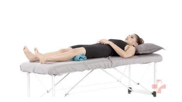 Quad sets (towel under knee)
Stop signs and precautions for the quad sets (towel under knee)
Before you start
- After a knee fracture or knee surgery, including a knee replacement, follow your surgeon's or physio's plan, and start this exercise only when they have cleared you.
When to stop and get help
- Your knee pain goes up sharply.
- Your calf or thigh is getting more swollen, or becomes warm, tender, red or darker than usual, or has a new throbbing or cramping pain. This can be a blood clot, especially after an operation, an injury, time in a cast, boot or brace, or a spell of being much less mobile than usual, and after an operation it can also be an infection. Stop and contact your medical team or get medical advice the same day. If you are also short of breath or have chest pain, call emergency services.
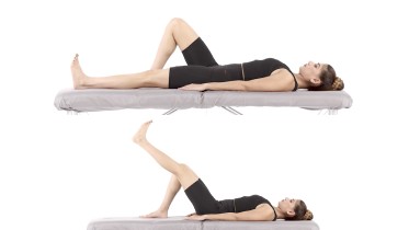 Straight leg raise
Stop signs and precautions for the straight leg raise
Before you start
- After a knee fracture or knee surgery, including a knee replacement, follow your surgeon's or physio's plan, and start this exercise only when they have cleared you.
- If you have had a hip replacement or other hip surgery, get the go-ahead from your surgeon or physio before you try this exercise. Some operations limit this kind of work for the first months, so the timing depends on your surgery. Lifting a straight leg is the heaviest way to use the muscles at the front of the hip, and after a periacetabular osteotomy a North American physiotherapy consensus panel asks for this exact movement, a straight leg lift while you are on your back, to be left out for 8 to 12 weeks.
- If you are pregnant and past the first three months, check with your physio before exercising flat on your back.
When to stop and get help
- Your lower back hurts.
- Knee pain increases during the set.
- You feel sharp pain in the front of the hip or groin.
- Your calf or thigh is getting more swollen, or becomes warm, tender, red or darker than usual, or has a new throbbing or cramping pain. This can be a blood clot, especially after an operation, an injury, time in a cast, boot or brace, or a spell of being much less mobile than usual, and after an operation it can also be an infection. Stop and contact your medical team or get medical advice the same day. If you are also short of breath or have chest pain, call emergency services.
- Your wound becomes more red, hot, swollen or painful, or starts to leak. Contact your medical team the same day.
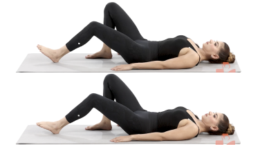 Hamstring sets (heel dig)
Stop signs and precautions for the hamstring sets (heel dig)
Before you start
- After a knee fracture or knee surgery, including a knee replacement, follow your surgeon's or physio's plan, and start this exercise only when they have cleared you.
- With a recent hamstring strain, even a still hold loads the injured muscle, so let your physio set when you start and how hard you press. The same applies after an ACL reconstruction that used a hamstring graft, where hamstring work is held back while the place the tendon was taken from heals, and the date comes from your surgeon's protocol. If you are pregnant and beyond the first trimester, ask your physio before you do this lying on your back.
- After a reconstruction of the posterolateral corner, the structures at the back of the outer side of the knee, leave this one out until your surgeon or physio tells you it is time. Digging the heel down to squeeze the back of the thigh is hamstring strengthening on its own, even though nothing moves and nothing resists you, and the protocol most widely followed holds hamstring strengthening on its own back for a minimum of four months, because the hamstring tendon on the outer side pulls on the fibular head, the small bone the graft is anchored into. So expect months rather than weeks there. If you have not had knee surgery, that wait does not apply to you.
- After an injury to the ligament at the back of the knee (the posterior cruciate ligament), the hamstrings pull the shin backward, which is the direction this ligament holds, so hamstring work is usually kept out of the early weeks. The knee stays close to straight in this hold, which is not where that pull is strongest, but your physio decides whether and when it fits, so do not start it on your own.
When to stop and get help
- Cramp in the back of the thigh, or a sudden sharp pull there. Stretch the leg out along the floor and rest.
- Knee pain that climbs sharply during or after the holds.
- Your calf or thigh is getting more swollen, or becomes warm, tender, red or darker than usual, or has a new throbbing or cramping pain. This can be a blood clot, especially after an operation, an injury, time in a cast, boot or brace, or a spell of being much less mobile than usual, and after an operation it can also be an infection. Stop and contact your medical team or get medical advice the same day. If you are also short of breath or have chest pain, call emergency services.
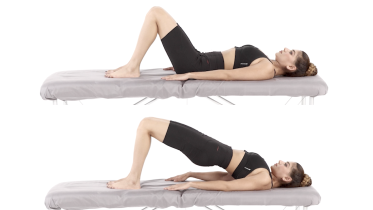 Glute bridge
Stop signs and precautions for the glute bridge
Before you start
- After hip, knee or back surgery, your surgeon's protocol decides when you can start bridges. Once you are past the first three months of pregnancy, check with your physio before exercising flat on your back.
- A bridge needs the knee bent to about 90 degrees before you lift, so if you have had knee surgery and been given a limit on how far the knee may bend, leave this one out and ask your surgeon or physio whether the bend it needs is allowed yet.
When to stop and get help
- You feel back pain rather than buttock work, even when you lift a little lower.
- Pain spreads down the leg or you get numbness or tingling.
- Your hamstrings (the back of the thighs) cramp. This is a common complaint with bridges, and bringing your feet a little closer to your bottom often eases it.
- Pain in the knee itself, or a knee that swells or is clearly more painful the next morning. The position holds the knee bent throughout, so after knee surgery stop and check with your surgeon or physio before you try it again.
Stage 2: The stretching program, plus loading with the foot down
The trial paired its strengthening with frequent stretching of the hamstrings, adductors, calves and quadriceps, which is why four stretches sit together here (Sauer 2022). Add the mini squat and the ball wall squat when stage 1 is comfortable, keeping them shallow at first so the kneecap and the fold under it are not squeezed hard. Leave the quadriceps stretch out if pulling the heel toward your bottom reproduces the pain at the front of the knee. Breathe steadily through every hold and never hold your breath, especially if you have high blood pressure. Four weeks of weekly physiotherapy review and then self-directed exercise is how the trial ran it, which is a reasonable shape to aim for.
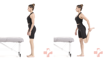 Standing quad stretch
Stop signs and precautions for the standing quad stretch
Before you start
- After a knee fracture or knee surgery, including a knee replacement, follow your surgeon's or physio's plan, and start this exercise only when they have cleared you.
When to stop and get help
- Sharp pain in the knee, especially at the front or behind the kneecap.
- You feel dizzy or unsteady. Hold on to support, or ask your physio about a lying version. If you faint while exercising, call emergency services, even if you feel fine again quickly.
- Tingling or numbness spreads down the front of the thigh.
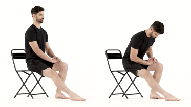 Seated hamstring stretch
Stop signs and precautions for the seated hamstring stretch
Before you start
- If you have had a hip replacement, get the go-ahead from your surgeon or physio before you try the seated hamstring stretch. Leaning forward from the hips while you sit bends the hip past a right angle, and many surgeons ask you to avoid that for the first months after some operations.
When to stop and get help
- Tingling, numbness, burning or pain running down the leg or into the foot.
- A sudden sharp pain, pop or snap at the back of your thigh. Stop straight away. If you then get large bruising spreading down the back of the thigh, a gap or dip you can feel, a stiff-legged walk, or clear weakness when you bend the knee, get assessed the same day at urgent care or an emergency department, as the tendon may have pulled away from the bone near the buttock.
- Pain in your lower back.
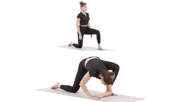 Half kneeling adductor stretch
Stop signs and precautions for the half kneeling adductor stretch
Before you start
- If you have had a hip replacement, get the go-ahead from your surgeon or physio before you try the half kneeling adductor stretch. Taking your elbows down beside the front foot folds that hip far beyond a right angle, and many surgeons ask you to avoid that for the first months after some operations. The kneeling hip is also taken back behind you, which some surgeons limit after a replacement done from the front of the hip.
- If you have had knee surgery or a knee replacement, check with your surgeon or physio before you kneel on that knee. Getting down to the floor and up again takes strength and balance, so have a sturdy chair next to you to lean on as you rise, and if you have fallen in the past year, ask your physio first.
- If you have high blood pressure, a heart condition, glaucoma or recent eye surgery, check with your doctor or physio first, keep breathing and come up slowly. If you have low bone density (osteoporosis), stay up on your hands with your back long rather than curling down to your elbows, and ask your physio whether this stretch suits you.
- If you are pregnant and have pain at the front or back of your pelvis, ask your physio first.
When to stop and get help
- Sharp or pinching pain deep in the groin or at the front of the hip.
- Pain, numbness or tingling that travels down the back of the leg as you straighten the knee.
- You feel dizzy, lightheaded or faint as you get up. Stay down or sit on the chair and wait for it to pass. If it does not pass within a minute or two, or it keeps happening, get medical advice the same day before you practice again. If you faint, or it does not settle within a few minutes of keeping still, call emergency services.
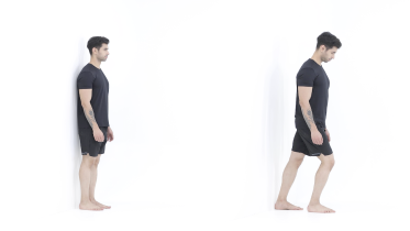 Calf stretch
Stop signs and precautions for the calf stretch
Before you start
- Calf stretching is often limited for the first months after an Achilles tendon repair, so follow your surgeon's and physio's instructions before you try this.
When to stop and get help
- Sharp pain in the Achilles tendon or at the back of the heel.
- Your calf or thigh becomes swollen, warm, tender, red or darker than usual, or has a throbbing or cramping pain that feels different from normal muscle ache. Stop and get medical advice the same day, as this can be a blood clot. If you are also short of breath or have chest pain, call emergency services.
- Tingling, numbness or burning in the foot or down the leg.
- Since the fracture or cast, the foot or ankle is getting more painful rather than less, the skin becomes very sensitive to light touch, or the foot changes color, temperature, swelling or sweating compared with the other one, or looks shiny. See your doctor or physio as soon as you can, within a few days at most, as this can be complex regional pain syndrome (CRPS) and early treatment may help.
- A sudden pop or snap at the back of your ankle or heel, a feeling that someone kicked you there, or sudden pain there after which you cannot push off or rise onto your toes on that leg. Stop, take the weight off that leg carefully and go to an urgent care center or emergency department the same day, even if you can still walk, as this can be a torn Achilles tendon.
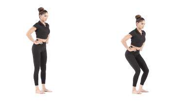 Mini squat
Stop signs and precautions for the mini squat
Before you start
- If you have had a hip replacement or other hip surgery, get the go-ahead from your surgeon or physio before you try mini squats. Some operations limit this kind of work for the first months, so the timing depends on your surgery.
- After a knee replacement, including a partial one, or other knee surgery, get the go-ahead from your surgeon or physio and ask how deep you may go. A squat bends the knee and loads it at the same time, so if you have been given a limit on how far the knee may bend, this is the exercise it applies to first. Keep it shallow and controlled until they say otherwise.
When to stop and get help
- Knee pain is sharp or keeps getting worse through the set.
- The knee swells, gives way or feels unstable afterwards.
- You feel dizzy, lightheaded, faint or sick, or the room seems to spin. Hold your support, sit down and get medical advice the same day before you practice again. If you faint, it does not settle within a few minutes of sitting still, your vision suddenly blurs or goes double, or you cannot stand or walk steadily, call emergency services.
- You feel unsteady enough that you might fall. Hold on to something firm and sit down.
- Your calf or thigh is getting more swollen, or becomes warm, tender, red or darker than usual, or has a new throbbing or cramping pain. This can be a blood clot, especially after an operation, an injury, time in a cast, boot or brace, or a spell of being much less mobile than usual, and after an operation it can also be an infection. Stop and contact your medical team or get medical advice the same day. If you are also short of breath or have chest pain, call emergency services.
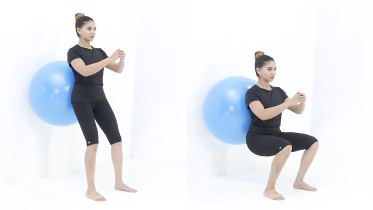 Ball wall squat
Stop signs and precautions for the ball wall squat
Before you start
- If you have had a hip or knee replacement, get the go-ahead from your surgeon or physio before you try the ball wall squat. Some operations limit this kind of work for the first months, so the timing depends on your surgery. Your surgical team will also tell you how far you are allowed to bend.
When to stop and get help
- Sharp pain in or around the kneecap, or knee pain that keeps building with each squat.
- Your knee gives way, or you lose control of the ball.
- Your lower back hurts more the lower you go.
- You feel dizzy, lightheaded, faint or sick, or the room seems to spin. Stop, sit down and get medical advice the same day before you exercise again. If you faint, it does not settle within a few minutes of sitting still, your vision suddenly blurs or goes double, or you cannot stand or walk steadily, call emergency services.
- Chest pain or pressure, dizziness or feeling faint, a racing or irregular heartbeat, or being far more out of breath than the effort should cause.
Stage 3: Back to stairs, chairs and single leg strength
When stage 2 feels easy and the knee is no worse the next morning, these take you back to the things daily life asks for. Use a low step to start with and lower slowly, with a rail or a wall to touch. The clamshell and side lying hip abduction are here because control of the whole leg affects how the kneecap sits, and poor kneecap tracking is one of the things the reviews ask you to address alongside the fold itself (Kuwabara 2021). If you have had a hip replacement, get the go-ahead from your surgeon or physio before you try the clamshell, the split squat and the step ups. Some operations limit this kind of work for the first months, so the timing depends on your surgery.
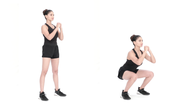 Squat
Stop signs and precautions for the squat
Before you start
- If you have had a hip or knee replacement, get the go-ahead from your surgeon or physio before you try squats. A deeper squat bends the hip well past a right angle, and many surgeons ask you to avoid that for the first months after some operations. Ask them how deep you may go.
When to stop and get help
- Sharp pain in the knee, hip or lower back, or pain that keeps building through the set.
- The knee gives way, locks or catches.
- The knee swells after the session.
- You feel dizzy, lightheaded, faint or sick, or the room seems to spin. Stop, sit down and get medical advice the same day before you exercise again. If you faint, it does not settle within a few minutes of sitting still, your vision suddenly blurs or goes double, or you cannot stand or walk steadily, call emergency services.
- Chest pain or pressure, dizziness or feeling faint, a racing or irregular heartbeat, or being far more out of breath than the effort should cause.
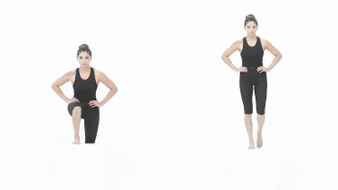 Step up
Stop signs and precautions for the step up
Before you start
- If you have had a hip or knee replacement, or other hip or knee surgery, get the go-ahead from your surgeon or physio before you try step ups. Some operations limit this kind of work for the first months, so the timing depends on your surgery. Your surgeon or physio will also tell you what step height is right for you.
- With a heart condition or high blood pressure, check with your doctor before you start.
- If you have fallen in the past year, have osteoporosis or feel unsteady on your feet, set the step beside a handrail, a kitchen counter or a sturdy chair back, keep one hand on it for every repetition, and ask your physio whether to start with step ups at all or to build up on a single stair first.
When to stop and get help
- You feel sharp pain in the knee or the knee gives way.
- Pain at the front of the knee builds with each rep.
- You feel dizzy, lightheaded, faint or sick, or the room seems to spin. Hold your support, sit down and get medical advice the same day before you practice again. If you faint, it does not settle within a few minutes of sitting still, your vision suddenly blurs or goes double, or you cannot stand or walk steadily, call emergency services.
- You feel unsteady enough that you might fall. Stop and sit down.
- Chest pain or pressure, dizziness or feeling faint, a racing or irregular heartbeat, or being far more out of breath than the effort should cause.
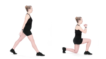 Split squat
Stop signs and precautions for the split squat
Before you start
- If you have had a hip or knee replacement, or other hip or knee surgery, get the go-ahead from your surgeon or physio before you try split squats. Some operations limit this kind of work for the first months, so the timing depends on your surgery.
- If you are pregnant, check with your midwife or physio before you start.
- With a heart condition or high blood pressure, check with your doctor before you start.
- If you have fallen in the past year, have osteoporosis or feel unsteady on your feet, set up beside a kitchen counter, a handrail or a sturdy chair back, keep one hand on it for every repetition, and ask your physio whether to start with split squats at all or to build up with both feet side by side first.
When to stop and get help
- You feel sharp pain in the knee, hip or groin, or the knee gives way.
- Knee pain builds with each rep, or is clearly worse the next morning.
- You feel dizzy, lightheaded, faint or sick, or the room seems to spin. Stop, sit down and get medical advice the same day before you exercise again. If you faint, it does not settle within a few minutes of sitting still, your vision suddenly blurs or goes double, or you cannot stand or walk steadily, call emergency services.
- Chest pain or pressure, dizziness or feeling faint, a racing or irregular heartbeat, or being far more out of breath than the effort should cause.
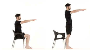 Sit to stand
Stop signs and precautions for the sit to stand
Before you start
- After a knee fracture or knee surgery, including a knee replacement, follow your surgeon's or physio's plan, and start this exercise only when they have cleared you.
- If you have had a hip replacement or other hip surgery, get the go-ahead from your surgeon or physio before you try sit to stand. Standing up from a low seat bends the hip past a right angle, and many surgeons ask you to avoid that for the first months after some operations. Ask them how high your seat should be. If you are pregnant, check with your midwife or physio before you start.
- Set up before the first stand: use a chair that will not slide, ideally with its back against a wall, and have someone nearby, or set the chair beside a kitchen counter you can reach, if you are not sure of your balance or you have fallen in the past year. Wear flat shoes that fit well and grip the floor, not socks, tights or bare feet.
When to stop and get help
- You feel dizzy, lightheaded or faint as you get up. Sit back down and wait for it to pass. If it does not pass within a minute or two, or it keeps happening, get medical advice the same day before you practice again. If you faint, or it does not settle within a few minutes of keeping still, call emergency services.
- Sharp pain in the knee, hip or lower back.
- Your knee gives way or you feel you might fall.
- Chest pain or pressure, dizziness or feeling faint, a racing or irregular heartbeat, or being far more out of breath than the effort should cause.
- You are pregnant and notice bleeding or fluid leaking from your vagina, regular painful tightenings, tummy pain that does not settle when you rest or comes with lower back pain, or a change in your baby's usual movements, such as moving less than usual. Stop and contact your midwife or maternity unit straight away. Do the same for a severe headache, problems with your vision such as blurring or flashing lights, pain just below your ribs, or sudden swelling of your face, hands or feet, which can be signs of pre-eclampsia. Call emergency services if the bleeding is heavy, the tummy pain is severe, bleeding or tummy pain comes with pain in your shoulder or feeling sick, faint or dizzy, or a headache comes on suddenly and is very severe.
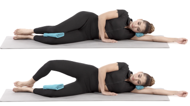 Clamshell
Stop signs and precautions for the clamshell
Before you start
- If you have had a hip replacement or other hip surgery, get the go-ahead from your surgeon or physio before you try clamshells. Opening the knee turns the thigh outward, and many surgeons ask you to avoid that for the first months after some operations. Lying on your side may also be limited in that time.
When to stop and get help
- You feel sharp or pinching pain in the hip or groin.
- Pain spreads into the leg, or you notice numbness or tingling.
- Pain on the outside of your hip builds during the set or is worse the next morning.
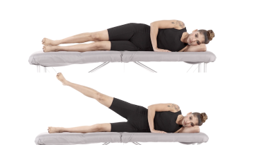 Side lying hip abduction
Stop signs and precautions for the side lying hip abduction
Before you start
- If you have had a hip replacement or other hip surgery, get the go-ahead from your surgeon or physio before you try side-lying hip abduction. Some operations limit this kind of work for the first months, so the timing depends on your surgery. With some surgical approaches, side leg lifts wait until the tissues at the side of the hip have healed.
When to stop and get help
- You feel sharp pain in the side of the hip, the groin or the lower back.
- Pain spreads down the leg or you get numbness or tingling.
Keeping moving between sessions
The trial's physiotherapy arm included a leg press, a stationary bike and an elliptical machine alongside the exercises above (Sauer 2022). We have no exercise page for any of the three, so they are worth mentioning here rather than leaving out.
If you have access to a bike or an elliptical, they are a reasonable way to keep the knee moving and the thigh working between strength sessions. Set the saddle high enough that the knee does not fold up sharply at the top of the pedal stroke, because holding the knee deeply bent for long spells is one of the things the reviews name as a provoker. Start with a short spin rather than a long one and judge it by the next morning.
Does conservative treatment work, and why do the sources disagree?
Three readings of the same question sit in the literature, and at first glance they do not reconcile. The randomized trial found physiotherapy equal to surgery at six months, with both groups beating the threshold for a meaningful change (Sauer 2022). Camanho reported 63 patients given 90 days of strengthening, flexibility work and activity modification, of whom 55 improved, 6 of those later had symptoms return that were never disabling, and 8 went on to arthroscopy (Camanho 2010). Schindler's review of the field, however, concludes that symptomatic plicae may initially be treated with physiotherapy and structured exercise but that success rates are generally low (Schindler 2014).
We read all three to see whether one of them settles it, and the useful finding is that they are not quite answering the same question.
Schindler's paper pooled 23 studies of plica excision, so the meta-analysis in it measured surgery, not exercise. The sentence about conservative success rates being generally low is the author's conclusion drawn from the wider literature, not a pooled result, and the same conclusion goes on to reserve excision for recalcitrant cases and for a fold that has undergone irreversible change. Camanho's series was not restricted to this fold. The commonest complaint in those 63 patients was pain above the kneecap, and the examination finding was pain on compressing the pouch above the kneecap, which points higher in the joint than the medial fold this page is about. The paper never reports how many of the 63 were medial, so it is a series of mixed folds rather than a comparator for this one, and its program was built around terminal knee extension. Sauer's trial is the one that studied the medial fold head to head, and it is the only randomized one.
So the honest summary is this. Exercise is the reasonable first step and all three sources say so. For the medial fold at six months, a trial found it matched an operation. What nobody has established is the long-run success rate of conservative care, and we are not going to invent one by picking a favorite source.
Severity is probably part of why the figures scatter, and one study gives you something to act on. In a series of 117 patients all given 3 months of conservative treatment, 76 did not improve and went to surgery. The ones who failed had a larger fold on MRI, 1.6 mm on average against 0.8 mm, more contact between the fold and the cartilage, more pain on the inner side as the knee moved from bending to straightening, and more snapping (Blanke 2021). Two of those four are measurements you cannot take at home. The other two you can report, and they are a reason to be reassessed rather than to keep grinding away alone. Worth knowing as well: the 41 who did get better without surgery ended up with scores as good as the surgical group's, so succeeding with exercise is not a second-best outcome.
What the operation is, and when it comes up
Arthroscopic resection means trimming the fold out through two small cuts, usually as a day case. Schindler's review pooled 23 studies and 969 patients after open or arthroscopic excision, mean age 25, with trauma considered the cause in 57% and a mean follow-up of 27.5 months: 64% were symptom free, 26% improved and 10% were failures (Schindler 2014). At 10 years, a series of 52 knees in 50 patients reported Lysholm scores rising from 52 to 80, with worse results where cartilage damage was present as well (Paczesny 2021).
Those are reasonable numbers and they are not a reason to skip the exercises, because the trial compared the two head to head and found no difference at six months. We do not set rehabilitation after this operation. If you are having it or have had it, the timings for weight bearing, range of movement and return to sport come from your surgical team.
When to see a physio or doctor
See a physio or doctor if the knee keeps catching, if focal pain on the inner side has not settled after a few weeks of changing what provokes it, or if you are not sure whether this is the fold, the meniscus or the kneecap joint. That last one is worth an appointment on its own, because the trial's authors found this diagnosis was wrong in both directions more often than not. Given that, an examination is worth more than another scan.
If you have done a properly supervised program for 3 months and the pain and snapping are unchanged, that is the point at which the literature starts talking about surgery, and it is a fair thing to raise.
For any of the warning signs below, get help within the time given there.
For physiotherapists
Sauer and colleagues 2022 is the only randomized trial. Its physiotherapy arm was quadriceps strengthening by quadriceps set, straight leg raise, leg press, stationary bike and elliptical, a frequent stretching program for hamstrings, adductors, gastrocnemius and quadriceps, avoidance of activity inducing plica irritation, and weekly physiotherapist review for 4 weeks followed by self-directed work. Note the tension in their own wording: they list a quadriceps set and a straight leg raise, both open chain, and state that open chain exercises that eventually cause plica irritation were avoided. The qualifier may resolve it, and the paragraph is worth reading before you build a protocol on it.
For the examination, Kuwabara and Fredericson describe the Stutter and Hughston tests and require both to be positive. Stubbings and Smith 2014 give the accuracy figures and argue for changing the reference standard away from arthroscopic appearance. Faber 2025 is the survey quantifying how divided the surgical community is.
Blanke 2021 gives the predictors of conservative failure, two radiological and two clinical, and also reports that the 41 of 117 who succeeded conservatively finished with a Lysholm of 96.25 against 95.93 in the operated group, with no difference in return to sport or Tegner. Hufeland 2019 is a prospective case series of 35 patients and 38 knees, mean age 16.2 and 73.7% female, with no control group, which the authors state.
On the conservative success disagreement, which earlier drafts carried as three irreconcilable readings: clinical review read all three and the scatter is partly a scope difference, not only an evidence one. Schindler's meta-analysis pooled 23 excision studies, so the "success rates are generally low" sentence is his conclusion from the wider literature rather than a pooled estimate, and it sits beside a recommendation to try physiotherapy first. Camanho's 63 patients presented mainly with suprapatellar pain and were examined by compression of the suprapatellar pouch, so that series is weighted toward the folds above the kneecap. It reports no breakdown by fold type at all, which is why clinical review 2 took out the earlier claim that most of the 63 were not medial: the paper does not support it, and Camanho's own terminology follows Iino's supra-, mid- and infrapatellar scheme, in which the midpatellar fold is the medial patellar plica. The protocol there was built on terminal knee extension, which this program does not copy because the trial's was not. Sauer is the only randomized trial and the only one of the three restricted to the medial fold. The page now says exercise first, matched surgery at six months for the medial fold, and no established long-run conservative success rate, rather than presenting the three as a stand-off.
Warning signs
Get emergency help now
- Emergency: the kneecap has slipped out of place, even if it went back by itself. If it is still out of place, go to an emergency department straight away, and do not drive yourself. If it went back by itself, get medical advice the same day.
- Your foot became weak or numb at the moment of the knee injury or in the hours after it, for example your foot drags, catches on the ground or slaps down when you walk (foot drop), you cannot pull your foot or toes upward, or the top of your foot has lost feeling. Go to an emergency department straight away. Do not drive yourself: ask someone to drive you or call an ambulance. The nerve that lifts the foot wraps round the bone on the outer side of the knee, and the same force that injures the knee can stretch it. A foot that has been weak or numb from that moment goes with the more severe injuries, where more than one ligament has been torn or the whole knee has been out of place, and in those the artery behind the knee has to be checked on the day. So this one is not watched at home to see whether it gets worse.
- Emergency: you are still growing, you have pain that wakes you at night, pain at rest that keeps getting worse, or a lump or swelling around the knee or thigh that keeps growing, and you also have a rash that does not fade when you press a glass on it. Call emergency services or go to an emergency department straight away. Without the rash, follow the line on the same symptoms further down this list, which gives you a day or two.
- Emergency: a sudden sharp pain, pop or snap at the kneecap or just above or below it while jumping, landing or sprinting, in someone who is still growing, and then the knee cannot be straightened, the leg cannot be lifted out straight or no weight can be put on it, or the knee swells quickly. Keep the weight off the leg and go to an emergency department straight away. In a growing knee this is a break where bone or cartilage pulls away at one end of the extensor mechanism rather than a torn tendon: either the bump at the top of the shin (tibial tubercle avulsion fracture) or the bottom of the kneecap (patellar sleeve fracture). The sleeve one can look almost normal on a first X-ray, so a child who still cannot straighten the knee afterwards needs to be seen again rather than reassured. This is not the catching or momentary giving way of a plica.
Get help today
- The pain started with a fall, a twist or a blow to the knee, or you cannot put weight on the leg. Get medical advice the same day. If the knee has changed shape, swelled up very quickly or you felt a pop or snap, or your foot turns cold, pale or blue or you have tingling or no feeling in the leg or foot, go to an emergency department straight away, and do not drive yourself.
- The knee is hot, red and swollen, or you have knee pain with a high temperature or feel hot, cold or shivery. Get medical advice the same day, and go to an emergency department if you feel very unwell. This can be a sign of infection in the joint, which needs treatment quickly.
- Same day: the knee is locked, which means it is stuck bent and you cannot fully straighten it, even after gently trying to move it, or you cannot bend or straighten the leg at all. Get assessed the same day by a doctor, at urgent care or at an emergency department. A torn piece of meniscus can get caught in the joint and block it, and when a tear like this can be repaired, it should be done as early as possible. A knee that catches for a moment and then frees itself can happen with a meniscus tear. That is not the same as a locked knee, but tell your physio or doctor about it.
- Same day: the knee keeps giving way under you, or it gave way after a recent injury, or it gives way and is also very painful or swollen. Get medical advice the same day. If it only happens now and then, with no recent injury and no swelling, it is not an emergency, but book an assessment with a physio or doctor in the next few days. Until it has been checked, leave out the exercises you do standing on one leg, hold the rail on stairs and take care on uneven ground, because a knee that gives way without warning is how people fall.
- The knee swells up with no clear reason, especially if you take medicine to thin your blood. Get medical advice the same day.
- A calf or thigh that is newly swollen or getting more swollen, warm, tender, red or darker than usual, has swollen veins that are sore to touch, or has a throbbing or cramping pain that does not fit your injury or condition, or that feels different from normal muscle soreness after exercise. This can be a sign of a blood clot (deep vein thrombosis), especially after surgery, an injury, time in a cast or boot, a long trip or a spell of being much less mobile than usual. Get medical advice the same day. If you are also short of breath or have chest pain, call emergency services.
- A leg or foot that is getting weaker, for example your foot drags, catches on the ground or slaps down when you walk (foot drop). Get medical advice the same day. If the weakness is getting worse by the hour, go to an emergency department. If the weakness started at the moment of your knee injury or in the hours after it, the emergency department line above applies instead.
- Same day: you are a teenager who is still growing and you have a limp, or pain in the hip, groin or thigh as well as the knee. Get an urgent appointment with a doctor the same day, and stay off sport and these exercises until you have been checked. If you cannot put weight on the leg, have a high temperature or feel very unwell, go to an emergency department. Some hip problems in growing teenagers, such as a slipped growth plate at the top of the thigh bone (slipped capital femoral epiphysis), are felt in the knee and need checking quickly.
- Pain at night or at rest that keeps getting worse. If you are being treated for cancer now, contact your cancer team the same day.
Book an appointment
- You are still growing and have pain that wakes you at night, pain at rest that keeps getting worse, or a lump or swelling around the knee or thigh that keeps growing. See a doctor within a day or two. Ask for an urgent appointment rather than the next routine one, and say what the pain does at night and whether a lump is getting bigger. Do the same if the pain comes with looking pale, unusual tiredness, fevers with no clear cause, bruising or bleeding easily, or weight loss. Bone pain that is worse at night, or a new lump, needs checking to rule out rarer causes such as a bone tumor or a blood cancer (leukemia).
- Pain at night or at rest that keeps getting worse. If you have had cancer, now or in the past, see your doctor sooner, within a day or two, and mention it, even if the pain is not getting worse. Do the same if you have knee pain and are losing weight without trying, even if it does not hurt at night.
- Pain at night or at rest that keeps getting worse. See your doctor within a few days.
Common questions
Is it a plica or a meniscus tear?
This is the question that brings most people to the name, and it cannot always be answered without an examination. Kuwabara and Fredericson write that plica syndromes are often missed because they are difficult to separate from meniscal tears, cartilage injuries and other lesions in the same area (Kuwabara 2021). Two things point toward the fold: pain you can localize to one small spot on the inner side of the front of the knee, rather than a diffuse ache around the kneecap, and snapping or catching through bending and straightening without a locked knee. A meniscus tear more often follows a twist, swells over hours, and can lock the knee so it will not fully straighten, which is a same day warning sign on this page. Our meniscus tear page covers that side.
Does everyone have a plica?
Most people do, which is the single most important fact on this page. Kuwabara and Fredericson report that anatomic studies indicate 90% of adults have one or more plicae in their knee joints, and that the vast majority are incidental and cause no symptoms (Kuwabara 2021). In a study of 3,889 knee joints in 3,563 Japanese patients having arthroscopic surgery, mean age 35, a medial plica was found in 79.9% (Nakayama 2011). Those were knees being operated on for other reasons rather than a general population sample, so read it as how often the fold is there, not how often it hurts. Being told you have a plica on a scan therefore tells you very little on its own.
How is plica syndrome diagnosed?
By examination and by ruling other things out, and the tests are not as good as you might hope. A systematic review pooling 7 studies and 492 knees found the medial plica examination test had a sensitivity of 0.90 and specificity of 0.89, ultrasound 0.90 and 0.83, and MRI 0.77 and 0.58, from literature of moderate methodological quality (Stubbings and Smith 2014). The MRI numbers deserve attention, because MRI is the test you are most likely to have had, and a specificity of 0.58 means it labels a lot of knees positive that do not have the problem. Kuwabara and Fredericson name two physical tests, the Stutter test and the Hughston test, and say both must be positive to support the diagnosis (Kuwabara 2021).
Do the exercises work for plica syndrome?
For many people yes, and this condition has better evidence for exercise than most of its neighbors, because it has an actual trial. In the randomized trial, 48 people with isolated medial plica syndrome had either arthroscopic resection of the fold or structured physiotherapy: at 6 months the two groups scored 82.3 and 83.1 on the Lysholm scale, with no significant difference, and both had improved by more than the 10.1 points the authors set as clinically meaningful (Sauer 2022). A separate series of 63 patients given 90 days of strengthening, flexibility work and activity modification reported 55 improved, though that series was not restricted to the fold this page is about: the commonest complaint was pain above the kneecap, the examination test was pressure on the pouch above it, and the paper never says how many of the 63 folds were medial ones (Camanho 2010). Set against that, the major review of the field concludes that success rates with conservative treatment are generally low, in a paper whose own meta-analysis pooled surgical results rather than exercise (Schindler 2014). The literature does not agree with itself here, so you are getting all three readings rather than the cheerful one on its own, and the section further down this page explains what separates them.
Will I need an operation?
Often not, and the decision is one for you and a surgeon rather than a website. What the trial found is that physiotherapy matched surgery at 6 months, then fell behind by 2 years, when the surgical group scored 89.7 and the physiotherapy group 74.6 on 33 of the 48 patients (Sauer 2022). The authors' own reading is that the benefit of physiotherapy is tied to continuing it, and they record that most patients said they had abandoned the exercise protocol after 3 to 6 months. If you are offered an operation, reasonable things to ask are what was seen on the scan, what else has been ruled out, and what happens if you give a properly supervised program three more months.
Does a knee brace help plica syndrome?
We cannot tell you, because the sources read for this page barely mention bracing, and the randomized trial did not use one. People ask this one constantly, which is worth saying plainly: the demand for an answer is not the same as an answer existing. Taping to improve how the kneecap tracks does appear in the reviews as part of conservative care, and a physio can try it and see whether it changes your pain (Kuwabara 2021). If a brace makes walking comfortable while you work through the program, that is a reasonable use of it rather than a treatment in its own right.
Which exercises should you avoid with plica syndrome?
The ones that reproduce the catch, which will be personal to you. The trial advised its physiotherapy group against activity that induced plica irritation, and the reviews say much the same: repetitive bending and straightening, and long spells with the knee held bent, are the usual provokers (Sauer 2022, Kuwabara 2021). In practice that often means cutting the depth of squats and lunges for a few weeks, breaking up long periods sitting with the knee folded up, and easing off a sudden increase in cycling or running mileage. Do not drop everything: the trial's benefit came from doing the program, not from resting.
References
- Sauer S, Karlsen G, Miller L, Storm JO. Medial Plica Syndrome of the Knee: Arthroscopic Plica Resection versus Structured Physiotherapy, A Randomized Controlled Trial. The Surgery Journal (New York). 2022;8(3):e249-e256. https://doi.org/10.1055/s-0042-1756183
- Schindler OS. 'The Sneaky Plica' revisited: morphology, pathophysiology and treatment of synovial plicae of the knee. Knee Surgery, Sports Traumatology, Arthroscopy. 2014;22(2):247-262. https://doi.org/10.1007/s00167-013-2368-4
- Nakayama A, Sugita T, Aizawa T, Takahashi A, Honma T. Incidence of medial plica in 3,889 knee joints in the Japanese population. Arthroscopy. 2011;27(11):1523-1527. https://doi.org/10.1016/j.arthro.2011.06.022
- Stubbings N, Smith T. Diagnostic test accuracy of clinical and radiological assessments for medial patella plica syndrome: a systematic review and meta-analysis. The Knee. 2014;21(2):486-490. https://doi.org/10.1016/j.knee.2013.11.001
- Faber S, Winkler PW, Henkelmann R, Diermeier T, Petersen W, Balke M, Metzlaff S, Colcuc S, Zimmermann G, Roessler PP, Niethammer TR. Treatment of a medial plica in the knee among German knee surgeons, The Plica Survey. Asia-Pacific Journal of Sports Medicine, Arthroscopy, Rehabilitation and Technology. 2025;40:18-22. https://doi.org/10.1016/j.asmart.2025.01.003
- Camanho GL. Treatment of pathological synovial plicae of the knee. Clinics (Sao Paulo). 2010;65(3):247-250. https://doi.org/10.1590/S1807-59322010000300002
- Blanke F, Oehler N, Al Aidarous H, Tischer T, Vogt S, Lenz R. Predictors for an unsuccessful conservative treatment of patients with medial patellar plica syndrome. Archives of Orthopaedic and Trauma Surgery. 2021;141(1):93-98. https://doi.org/10.1007/s00402-020-03646-6
- Hufeland M, Treder L, Kubo HK, Verde PE, Krauspe R, Patzer T. Symptomatic medial synovial plica of the knee joint: an underestimated pathology in young patients. Archives of Orthopaedic and Trauma Surgery. 2019;139(11):1625-1631. https://doi.org/10.1007/s00402-019-03249-w
- Paczesny L, Zabrzynski J, Kentzer R, Gryckiewicz S, Lewandowski B, Szwedowski D, Kruczynski J. A 10-Year Follow-up on Arthroscopic Medial Plica Syndrome Treatments with Special Reference to Related Cartilage Injuries. Cartilage. 2021;13(1_suppl):974S-983S. https://doi.org/10.1177/1947603519892310
- Kuwabara A, Fredericson M. Narrative: Review of Anterior Knee Pain Differential Diagnosis (Other than Patellofemoral Pain). Current Reviews in Musculoskeletal Medicine. 2021;14(3):232-238. https://doi.org/10.1007/s12178-021-09704-9
Written and checked by the PocketPhysio editorial team. Last updated 2026-11-06.
This page is general education, not a diagnosis or a personal treatment plan. See a physiotherapist or doctor for advice about your own situation.