
High ankle sprain: recovery and rehab exercises
Warning signs, and when to get help
What is a high ankle sprain?
Your two shin bones, the tibia and the fibula, run side by side down to the ankle and are bound together at the bottom by a set of ligaments called the syndesmosis. A high ankle sprain is an injury to that binding. OrthoInfo describes it as an injury to the ligaments and structures connecting the tibia and fibula. Syndesmotic is the word you will see on a clinic letter or a scan report.
Usually the foot is planted and the lower leg twists over it, or the foot is forced upward and outward. We see it after tackles, in ski boots, when a foot catches in turf, and from an awkward step off a curb. The sore spot sits above the ankle joint, not on the bony bump at the outside, and pressing along the front of the gap between the two bones usually finds it.
How is a high ankle sprain different from a normal sprain?
Most sprains are not this one. OrthoInfo puts about 90% of ankle sprains in the inversion group, where the foot rolls under and the ligaments on the outer side are stretched or torn. Those settle with the sort of program on our ankle sprain page.
The syndesmosis is a different job. It stops the two shin bones spreading apart while the talus, the bone they sit on, is driven up between them with every step. So the movements that hurt are the ones that push the bones apart: bending the ankle forward, and the foot turning outward. That is also why the two injuries need different rehab. A lateral sprain is rebuilt around inversion and eversion control, and this one is rebuilt around tolerating forward bend and rotation, which is why the band work below leaves resisted eversion until stage 2 and builds it slowly.
A high ankle sprain also feels worse than people expect. Push-off stays painful long after walking on the flat is fine. Stairs and hills demand forward bend, so they stay sore for weeks. The swelling is often unimpressive next to how much it hurts, which is what makes people assume the injury is minor.
How long does a high ankle sprain take to heal?
There is no single number, and the reason matters. If the joint is stable, this is rehabilitated without an operation and the timeline sits at the longer end of ankle sprain recovery. OrthoInfo describes its three phases as taking about 2 weeks for minor sprains and 6 to 12 weeks for more severe injuries, a figure given for ankle sprains as a group rather than for syndesmotic injuries alone.
If the joint is unstable, it is usually held with a screw or a suture button, and the timeline is then set by the operation and the protocol. Nobody should offer you a confident week count here. When the ISAKOS leg, ankle and foot committee surveyed 742 providers about syndesmotic injuries, return to play timelines varied widely between them, and most respondents used the same rehabilitation protocol regardless of how severe the injury was (Hunt and colleagues, 2022). A review of return to play after foot and ankle injuries puts it more carefully: some data support an earlier return to sport and higher function scores with flexible fixation than with a rigid screw (Allahabadi and colleagues, 2020).
The two ends of the published range sit a long way apart. One clinical viewpoint on an accelerated protocol after suture button fixation has athletes tested for return to play at four to six weeks, and the same authors put the usual time out of sport after a screw at three to six months (Voight and colleagues, 2024). We have left that protocol's day by day weight bearing and boot dates off this page on purpose. It is a viewpoint rather than a study, written for athletes who have a surgeon and a therapist on hand every day and for one type of flexible implant, and weight going through the leg sooner than your own surgeon allows is the mistake this page most wants you to avoid.
For a figure from an ordinary clinic population instead: among 46 adults treated for an unstable syndesmosis, average age about 41, average time to full weight bearing was 8 weeks after a suture button and 9.1 weeks after a screw (Naqvi and colleagues, 2012). That is nothing like the athlete timelines, and neither set of numbers is yours. Your surgeon's dates are.
Signs of a high ankle sprain, and the tests a clinician uses
Tenderness above the ankle joint, between and over the two shin bones, is the finding that points here. Pain on squeezing the calf higher up, pain when the foot is rotated outward, and pain with a deep forward bend of the ankle are the other three a clinician uses. None of them is a self-test, and we are not giving you a way to check at home, because the thing that matters most is whether the joint is stable, and that cannot be felt from the outside.
Imaging decides it. X-rays are taken to rule out a fracture, OrthoInfo notes that stress X-rays show ligament related instability, and an MRI is not required but may be ordered to assess a high ankle sprain. Mention any pain higher up the outside of your leg to whoever examines you, because the same twist can break the fibula up near the knee, and that break needs the whole lower leg on the X-ray, not the ankle alone. In a series of 70 adults with that injury pattern, the fibular fracture showed only on the side view of the lower leg in 24% of them (Kasper and colleagues, 2020). If you heard a crack at the time, cannot put weight on the leg, or the ankle has changed shape, that is an emergency department visit rather than a physio appointment.
How to use this program
Read the warning signs at the foot of this page before you start. If your ankle was operated on, follow the plan from your surgeon and physio instead of this one, and bring this page to them rather than the other way round. If you are in a boot or a brace, only put weight through the leg as much as your treating team has allowed.
Pick the stage that matches where you are today, and start at stage 1 if you are not sure. Move up when the current stage feels easy and the ankle settles well by the next morning. Each exercise page gives a typical starting dose, and as a rough guide many programs use 2 to 3 sets of 10 to 15 repetitions. Your physio will adjust the exercises and the numbers to suit you.
Breathe normally through every repetition rather than holding your breath, especially if you have high blood pressure. If you have fallen in the past year, feel unsteady on your feet, or take medicines that make you dizzy, keep a hand on a kitchen counter for the whole of the standing work, practice only when someone else is at home, and ask your doctor or physio about a falls check.
The pain rule for this injury is specific. Muscle fatigue and a mild ache that settles the same evening are fine. Pain felt in the gap between the two shin bones, at the front above the joint, means the load reached the healing ligaments and you have gone too far for today.
The exercise program
Stage 1: Settle it down and keep everything else working
For the first days to weeks, while you are in a boot, a cast, a brace or a wrap. If your leg is in a cast, the ankle exercises here are not open to you, so ask your fracture clinic or surgical team which movements you may do, and work the hip in the meantime, along with the toes if the cast leaves them free and your team is happy for you to move them. The syndesmosis is loaded by bending the ankle up and by the foot turning outward, so this stage stays away from both and leans on holds rather than end-range movement. Ankle pumps are done gently and within comfort, not pushed into a stretch. The exercise page shows them sitting with both feet flat on the floor, which presses the ball of the foot down as the heel lifts, so if you have been told to keep weight off the leg, do them one leg at a time, with the injured foot off the floor and that leg resting on a stool or a bed instead. The isometric holds ask the muscles to work without the joint moving, and the outward one is pressed lightly rather than hard, because a strong outward push is the direction that loads the injured ligaments. The outward hold also has a setup you only find out about when you open its page: it is done sitting on the floor with a soft ball between the outside of your foot and a wall. If you are on crutches, or getting down to the floor and back up again is not safe for you yet, do that hold sitting on a chair instead, with the outside of your foot pressed lightly against a wall or a heavy table leg and your heel resting on the floor. The glute bridge keeps the hip from switching off while you are limping, and it is the one exercise here that presses through your heels: if you have been told to keep all weight off the injured leg, leave it out until your treating team confirms that heel pressure is allowed, and add it once they do. The straight leg raise and the clamshell are the hip work that puts nothing at all through the foot, so they are what to use in the meantime, and they are also the two to work on while your leg is in a cast, with the toe curls added if your team has said the toes may move. If you are pregnant and past the first three months, check with your physio before exercising flat on your back. Many programs use 2 to 3 sets of 10 repetitions, or 5 to 10 holds of 5 to 10 seconds, once or twice a day. Your physio will adjust this. Only put weight through the leg as much as your treating team has allowed.
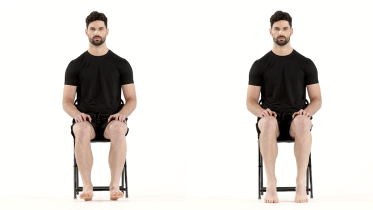 Ankle pumps
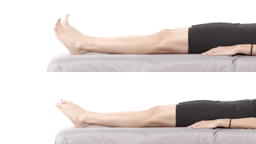 Toe curls
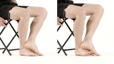 Isometric ankle dorsiflexion
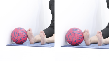 Isometric ankle eversion
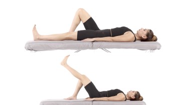 Straight leg raise
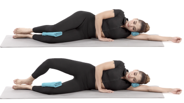 Clamshell
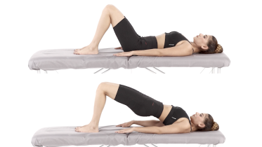 Glute bridge
Stage 2: Get the movement back and start band work
When the sharp pain has gone and your team is happy for you to move the ankle through range, often two to four weeks in but sometimes later. Circles and the alphabet take the ankle in every direction without weight through the foot. Both of those exercise pages ask for the biggest circles and the largest letters you can make, and this is the one place to read them loosely: keep them inside comfort and stop short of the end of the turn outward and of the bend upward, because those are the two directions that stress the injured ligaments. The four band directions then add resistance in a sitting position, and two of them are the ones to build slowly: the band that pulls your foot outward, and the band you pull your foot up against, since turning the foot out and bending the ankle up are the two movements that load this injury. Do the seated calf raise with no weight on your knees to begin with, even though the exercise page shows a dumbbell resting there, because anything you put on your knees travels down through the ankle, and keep the total inside what your team has allowed through the leg. Many programs use 2 to 3 sets of 10 to 15 with the band, most days while the band is light, dropping to 3 days a week with a day between once you move up to a heavier band. Your physio will adjust this. Stop a set if you get pain in the gap between the two shin bones rather than muscle fatigue.
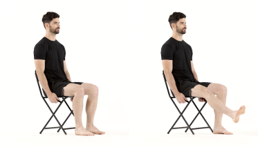 Ankle circles
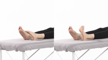 Ankle alphabet
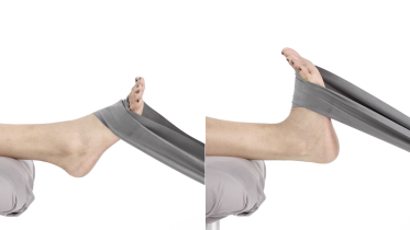 Ankle dorsiflexion with band
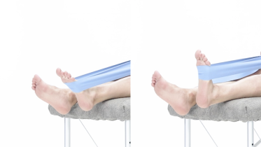 Ankle plantar flexion with band
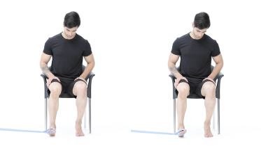 Ankle inversion with band
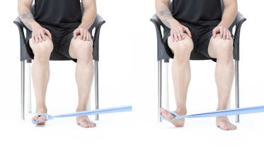 Ankle eversion with band
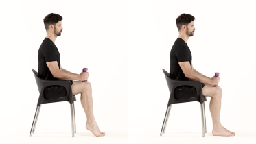 Seated calf raise
Stage 3: Load it standing, and get the calf back
Start this stage when you are walking without a limp in a normal shoe and the band work feels easy. Calf raises on both feet rebuild the push-off you lost in the boot. The two calf stretches and the half-kneeling stretch are for an ankle that still will not bend forward enough for stairs or a slope, and this is the one place where forward bend is worked on purpose rather than avoided, so build into it over weeks rather than forcing it. Take them in that order, and make the half-kneeling stretch the last thing you add in this stage. Start it only once the calf raises on both feet are comfortable for 2 to 3 sets of 10 to 15 and the two standing stretches have left the ankle no worse the next morning. It is the most loading thing in the stage: it drives your knee out past your toes with your body weight over a flat heel, which is close to the position a clinician uses to test a syndesmosis. How far it goes is set by how far you lean, so lean less than you think you can. Keep a chair or a counter beside you for getting down into half kneeling and back up, and if you cannot get down to the floor safely, stay with the two standing stretches and ask your physio for another way to work the forward bend. If your ankle was operated on, those three stretches are the ones to clear with your operating surgeon by name before you start them, because forward bend with your weight on the foot loads the screw or the suture button rather than the ankle in general. Balance goes from feet together to heel to toe to standing on the injured leg, with a counter within reach. Many programs use 2 to 3 sets of 10 to 15 for the calf raises, stretch holds of about 30 seconds repeated 2 to 3 times on each leg, and balance holds of 10 to 30 seconds. Strength work is often done 3 days a week with a day between, and balance work most days. Your physio will adjust this.
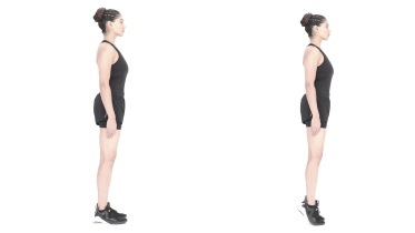 Calf raises
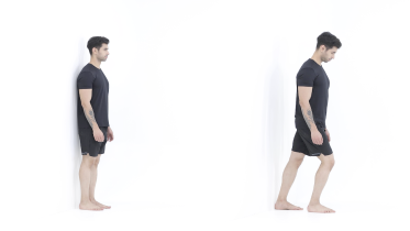 Calf stretch
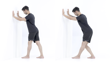 Soleus stretch
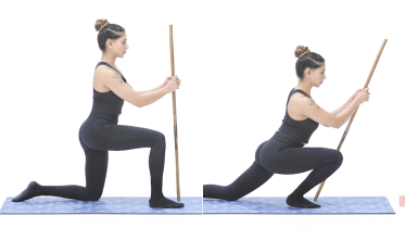 Half-kneeling ankle stretch
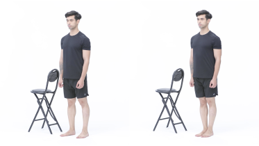 Standing with feet together
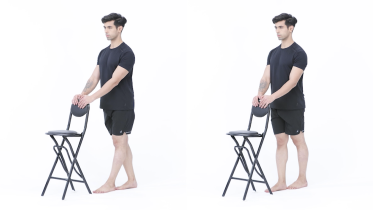 Tandem stance
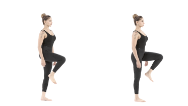 Single leg stance
Stage 4: Hopping, cutting and back to sport
For when you are through stage 3, the calf raises on both feet are comfortable at 2 to 3 sets of 10 to 15, and you are walking without a limp in a normal shoe. Single leg heel raises and step ups are the strength base, and they come first, because stage 3 only ever asked the calf to work on two feet. Build the single leg rise up over several sessions with a hand on a counter rather than finding out in one go how many you can do. The hopping drills then wait until you can rise onto the injured leg alone at least 10 times with the knee straight, within a few repetitions of what the other leg manages, and the ankle settles well the next morning. Pogo hops teach a stiff, quiet landing, the single leg hop and hold tests whether you can absorb it on one leg, and fast feet and skater hops bring in the side to side and turning loads that a high ankle sprain dislikes most. If your ankle was operated on, the whole of this stage waits for your operating surgeon's clearance rather than for the ankle feeling ready. Start with two sessions a week, well away from each other, 2 to 3 sets of 5 to 10 landings in each drill to begin with, and add volume before you add speed or direction changes. Your physio will adjust this. A physio can compare the two legs for strength and hopping before you go back to a cutting sport. Jumping and hopping also load the joints, the bones and the pelvic floor. If you have osteoporosis or have had a fall in the past year, ask your physio before you do any hopping. If you have had a hip or knee replacement, get the go-ahead from your surgeon before you try hopping. Many surgeons advise against jumping and hopping after a joint replacement for good, not just for the first months, because each landing sends high impact through the new joint. If you are pregnant, have had a baby in the past year, or leak urine when you jump, cough or sneeze, check with your midwife or physio before you start this stage. Stop the hopping and speak to your physio if urine leaks while you are doing it, or if you feel pressure or heaviness low in your pelvis. After having a baby, postnatal guidance usually leaves jumping and hopping until at least 3 months after the birth, once a pelvic floor check shows you are ready. With a heart condition or high blood pressure, check with your doctor before you start.
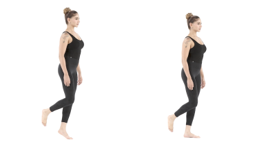 Single leg heel raise
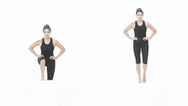 Step up
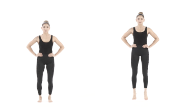 Pogo hops
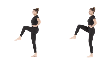 Single leg hop and hold
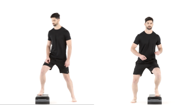 Fast feet side to side
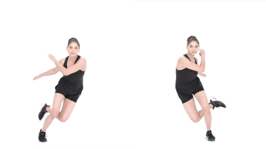 Skater hops
Taping, strapping and a brace: what they do and what they do not
Taping, strapping and a brace all do the same thing: they make the ankle feel steadier and more comfortable while you load it. OrthoInfo describes taping or bracing being used during an early return to sport or work. We have not found a trial showing that any of them changes how a syndesmotic injury heals, so the honest framing is support, not treatment.
That matters because a strapping is good enough to let you train on an ankle that is not ready. If tape is the only reason you can play, the strength is not there yet, and the hop tests in stage 4 are a better guide than how the tape feels. A boot is different again: it is there to stop the movement that stresses the injury, and when it comes off is your team's decision, not a comfort judgment.
When a high ankle sprain needs surgery
OrthoInfo lists high ankle sprains with instability of the ankle syndesmosis among the situations where surgery is considered, along with injuries that have not responded to non-surgical care and persistent instability and pain after months of rehabilitation. The operation holds the two shin bones at the right distance while the ligaments knit, using either a rigid screw or a flexible suture button construct.
Whether an injury counts as unstable is measured, not judged by how much it hurts. The surgeons in the ISAKOS survey most often named widening of the syndesmosis of more than 2 mm on X-ray, a torn anterior ligament along with injury to the posterior ligament or the deltoid on MRI, and widening of the joint seen during arthroscopy. Flexible devices were the preferred construct for 47% of them and screws for 30% (Hunt and colleagues, 2022). If a screw is what you are having, the same authors who wrote the accelerated protocol note that a screw may need a second procedure to remove it and carries a risk of breaking during rehabilitation (Voight and colleagues, 2024), so ask what the plan is for taking it out.
If you have been offered an operation, the questions worth asking are which fixation is being used, whether anything is coming out later, how long you are off the leg, and what the protocol says about running. Protocols differ between surgeons more than patients expect, which is what the ISAKOS survey measured.
After fixation for an unstable syndesmosis
This page does not stage post-operative rehab, because the weight bearing dates, the range limits and the point at which impact starts belong to the surgeon who did the operation. What this page can do is tell you what to watch for and where the exercises fit once you are cleared.
Two movements load the fixation itself rather than the ankle in general: bending the ankle forward while you are standing on it, and the foot turning outward. Those two, and the date weight can go through the leg, are the three things to get from your surgeon in words you can repeat, because they are what differs most from one surgeon to the next. The ISAKOS survey found considerable variation between respondents in time to weight bearing, running and full participation (Hunt and colleagues, 2022). That is also why the stage 3 calf stretches, which deliberately push the forward bend, and all of stage 4 need clearing with your surgeon by name rather than being started because the ankle feels ready.
The warning signs below include two written for you specifically: a wound that is not behaving, and the signs that the fixation may be losing position. Both go to your surgical team the same day. Our ankle fracture rehab page covers the boot, weight bearing and swelling material in more detail, because most of that is the same whether what was fixed was a bone or a ligament complex.
Getting back to running and sport
Impact is reintroduced on strength and symmetry, not on the calendar. The practical gates are walking without a limp in a normal shoe, rising onto the injured leg alone at least 10 times with the knee straight and within a few repetitions of what the other leg manages, and an ankle that is no worse the next morning after a session. Then hopping, then straight line running, then turns.
Our return to running protocols guide covers the build-up in detail. If the ankle keeps giving way or you have had several sprains, read chronic ankle instability as well, and if the pain has moved to the outer side behind the bony bump, peroneal tendinopathy is a common companion to an ankle that has been guarded for weeks.
When to see a physio or doctor
Get it looked at early rather than late. Any new ankle injury where you cannot put weight on the leg, heard a crack, or have a swollen ankle that has changed shape needs assessment that day. Beyond that, see a physio or doctor if you are not clearly improving over two to three weeks, if push-off is still painful at three months, or if the ankle feels unsteady on uneven ground.
Check with your doctor before you start if you have a heart or lung condition, or blood pressure that is not under control. If you have diabetes or reduced feeling in your feet, have the foot checked rather than managing a new ankle injury at home.
For physiotherapists
The useful distinction at assessment is stable against unstable, and the page is written to keep that question open rather than to settle it. Load the forward bend and rotation deliberately rather than avoiding them indefinitely, since tolerance to both is what the return to sport actually requires. Side to side comparison of single leg heel raise endurance, weight bearing dorsiflexion range and a hop battery gives something to track, and the ISAKOS survey is worth reading before quoting any return to play week count to a patient.
Warning signs
Get emergency help now
- Emergency: sudden, severe pain in your lower leg that is much worse than you would expect, especially after an injury or with a plaster cast or tight bandage on the leg. It may come with a tight, swollen leg, pain that gets worse when the muscle is stretched, numbness, pins and needles or weakness. Call emergency services or go to an emergency department straight away, and do not drive yourself. This can be acute compartment syndrome, which needs urgent surgery.
- Emergency: while you are in a cast or boot, your toes turn blue, white or pale, or feel cold. Do not wait to see if raising your leg helps. Go to an emergency department straight away, and do not drive yourself. The cast or boot may be cutting off the blood supply, or pressure may be building up inside the leg (compartment syndrome), and both need checking quickly.
- Part of your foot or a toe turns cold, pale, blue or black. Call emergency services or go to an emergency department straight away, and do not drive yourself, as the blood supply to the foot may be blocked.
Get help now, day or night
- In a cast or boot, the pain gets worse, your toes feel numb, tingle or burn or look more swollen, or the cast or boot feels too tight. Stop, raise your foot to about hip level and no higher, keep moving your toes and contact your surgical team or fracture clinic or get medical advice straight away. If you wear a boot, loosen the straps but keep it on. Do not prop the foot higher than that and do not put ice on it, because if pressure is building up inside the lower leg both of those cut down the blood reaching your foot. If you cannot move your toes, the pain is severe even after painkillers, or the numbness, tingling, burning or swelling has not settled within 30 minutes of raising your foot, go to your fracture clinic or an emergency department straight away, and do not drive yourself.
- Your cast or boot rubs painfully, or there is a blister-like soreness under it, or there is a bad smell, discharge or liquid coming from under the cast, or something has dropped down inside it. Contact your fracture clinic, plaster room or surgical team straight away, and out of hours go to an emergency department rather than waiting for the morning. Skin under a cast can break down where you cannot see it.
Get help today
- Your ankle suddenly becomes much more painful, or you can no longer put weight on it when you could before, for example after a slip, a trip or a twist. Keep your weight off it and get it checked the same day at your fracture clinic or an emergency department. If the ankle looks out of shape or sits at an odd angle, or a bone is showing through the skin, go to an emergency department straight away, and do not drive yourself.
- New pain or tenderness high on the outside of your leg, up near the knee, after the twist that hurt your ankle, whether or not you can walk on it. The same twisting force that tears these ligaments can also break the fibula high up, and that break is easy to miss on an X-ray that stops at the ankle. Keep your weight off the leg and get it assessed the same day at an urgent care center, a fracture clinic or an emergency department.
- Since your operation, your wound is oozing, leaking or has pus coming out of it, or its edges start to come apart, or your ankle is tender, swollen, painful or red and it is not getting better or is getting worse, or you have a high temperature or feel hot and cold or shivery. Redness can be harder to see on brown or black skin. Contact your surgical team the same day, or get urgent medical advice if you cannot reach them. These can be signs of an infection in the wound or the ankle joint. If you feel very unwell, go to an emergency department. If you have a very high or very low temperature, shivering you cannot control, you are breathing very fast or finding it hard to breathe, you are confused or your speech is slurred, your skin, lips or tongue look blue, gray, pale or blotchy (on brown or black skin this can be easier to see on the palms of the hands or soles of the feet), or you have a rash that does not fade when you press it, call emergency services, as this can be sepsis.
- Since your operation, your ankle pain is getting worse rather than settling as the weeks go on, including in the weeks after a screw has been taken out, you get a new pain between the two shin bones or on the inner side of the ankle, you feel a crack, snap or sudden give in the operated ankle, the ankle starts to look out of line or to sit differently in the boot or cast, a boot or cast that fitted starts to feel loose and roomy, you can feel a new hard or tender lump on the outer side of the ankle where the metal sits, or the ankle feels unstable or gives way when you put weight on it. A screw, plate or suture button holds the two shin bones in the right position while the ligaments knit, and that position can be lost without any new injury and without a snap, and a screw can loosen or break while you are rehabilitating. It is put right more easily when it is found early. Stop the exercises, keep your weight off that leg, keep the boot or cast on if you are still in one, and contact your surgical team or fracture clinic for an X-ray the same day. If the ankle looks clearly out of shape, or you cannot stand on it and cannot reach anyone, go to an urgent care center or an emergency department.
- A calf or thigh that is newly swollen or getting more swollen, warm, tender, red or darker than usual, has swollen veins that are sore to touch, or has a throbbing or cramping pain that does not fit your injury or condition, or that feels different from normal muscle soreness after exercise. This can be a sign of a blood clot (deep vein thrombosis), especially after surgery, an injury, time in a cast or boot, a long trip or a spell of being much less mobile than usual. Get medical advice the same day. If you are also short of breath or have chest pain, call emergency services.
- A leg or foot that is getting weaker, for example your foot drags, catches on the ground or slaps down when you walk (foot drop). Get medical advice the same day. If the weakness is getting worse by the hour, go to an emergency department.
- Same day: your cast feels too loose or uncomfortable, cracks, softens, gets wet or itches badly. Ring your surgical team, fracture clinic or plaster room and ask to be seen, and go to an emergency department if they cannot see you that day. A cast or boot that has gone loose or soft may no longer be holding your ankle in the position it was set in, so keep your weight off that leg until you have been seen, and do not cut, pad or adjust it yourself.
Book an appointment
- Since the injury, operation or boot, your foot or ankle is getting more painful rather than less, often with a burning pain that feels far worse than the injury, the skin becomes very sensitive to light touch, bedclothes or a change in temperature, or the foot changes color, temperature, swelling or sweating compared with the other one, or looks shiny. See your doctor or physio as soon as you can, within a few days at most, as this can be complex regional pain syndrome (CRPS) and early treatment may help.
- Your ankle is still painful at the front of the joint when you push off, still will not bend forward as far as the other side, or still feels unsteady on uneven ground, 3 months after the injury. This is not an emergency, but ask your doctor or physio for a review in the next week or two, because a syndesmotic injury that was managed as a simple sprain sometimes turns out to have been unstable, and that is answered by imaging rather than by more exercises.
Common questions
How long does a high ankle sprain take to heal?
It depends on whether the joint was stable, and anyone who gives you one number is guessing. For ankle sprains in general, OrthoInfo from the American Academy of Orthopaedic Surgeons puts the rehabilitation phases at about 2 weeks for minor sprains and 6 to 12 weeks for more severe injuries, and that figure covers ankle sprains as a group rather than syndesmotic ones specifically. For unstable injuries that were fixed surgically, there is no settled answer: when the ISAKOS leg, ankle and foot committee surveyed 742 providers, most of them surgeons, return to play timelines varied widely, and varied most according to whether the deltoid ligament on the inside of the ankle was injured too (Hunt and colleagues, 2022). Your own timeline comes from your treating team, not from a page.
What is the difference between a high ankle sprain and a normal ankle sprain?
The injury sits above the ankle joint rather than on the outside of it. A normal sprain tears the ligaments on the outer side when the foot rolls under, and OrthoInfo puts about 90% of ankle sprains in that group. A high ankle sprain injures the ligaments between the two shin bones, which is why the sore spot is higher up and why it hurts when the foot is turned outward or the ankle is bent forward. It is also why push-off and stairs stay sore for longer: every step pulls on the structure that was injured.
Does a high ankle sprain need surgery?
Only if the joint is unstable. OrthoInfo lists high ankle sprains with instability of the ankle syndesmosis among the reasons surgery is considered, alongside sprains that have not settled after months of rehabilitation. A stable injury is treated without an operation. The decision rests on examination and imaging, sometimes with stress X-rays, and it is a surgeon's call rather than something you can judge from how much it hurts.
Does taping or strapping help a high ankle sprain?
It can make walking and early sport more comfortable, and it does not speed up ligament healing. OrthoInfo describes taping or bracing during the early return to sport or work as part of normal management of ankle sprains. We have found no trial that shows taping or strapping changes how a high ankle sprain heals, so treat it as support while you build strength rather than as treatment. If a strapping lets you train on an ankle that still cannot take the load, it is working against you.
Can I walk on a high ankle sprain?
Follow what your treating team told you, because that instruction is based on whether the joint is stable. Some people are allowed to walk in a boot straight away and some are kept off the leg. Until you have been seen, the NHS advice for a sprain that stops you putting any weight on it or walking more than a few steps is to contact NHS 111 or go to an urgent care service, because an injury that severe needs assessment. Do not test it by trying to walk further.
What exercises should I avoid with a high ankle sprain?
Early on, anything that forces the ankle up into a deep forward bend or turns the foot outward against resistance. That means deep lunges, a loaded calf stretch on a step and hard resisted eversion are left until later. Jumping, hopping and anything with a turn in it come last, not first. None of these movements is banned forever, which is why the program works up to them. If a movement produces pain in the gap between the two shin bones rather than muscle effort, it has come too soon.
How do I know if my high ankle sprain is not healing?
Watch the push-off and the forward bend rather than the swelling. Pain at the front of the joint when you push off, or an ankle that still will not bend forward as far as the other side three months after the injury, both deserve a review. OrthoInfo also notes that an MRI may be considered when symptoms continue beyond 6 to 8 weeks. A high ankle sprain that was treated as a simple sprain and is not settling is one of the injuries worth going back about, because an unstable syndesmosis that was missed does not improve with more rehab.
Can a high ankle sprain cause long term problems?
It can, which is the honest answer. OrthoInfo says repeated ankle sprains can lead to chronic pain, instability and arthritis, and that incomplete rehabilitation is the most common cause of ongoing instability. The syndesmosis also sits right at the ankle joint, so an injury that heals in a poor position is one of the routes to ankle arthritis later. Finishing the strength and balance work, rather than stopping when the pain goes, is the part you control.
References
- American Academy of Orthopaedic Surgeons. Sprained Ankle. OrthoInfo. https://www.orthoinfo.org/en/diseases--conditions/sprained-ankle/
- Hunt KJ, Bartolomei J, Challa SC, McCormick JJ, D'Hooghe P, Tuffiash M, Amendola A. Significant variations in surgical construct and return to sport protocols with syndesmotic injuries: an ISAKOS global perspective. Journal of ISAKOS. 2022;7(1):13-18. https://doi.org/10.1016/j.jisako.2021.10.005
- Voight ML, Norman C, Wilk KE, Lucas M, Wolfe C. Management of High Ankle Sprains Utilizing the Tightrope Surgical Procedure: A Novel Approach for a Rapid Return to Play. International Journal of Sports Physical Therapy. 2024;19(5):513-521. https://doi.org/10.26603/001c.116862
- Naqvi GA, Cunningham P, Lynch B, Galvin R, Awan N. Fixation of ankle syndesmotic injuries: comparison of tightrope fixation and syndesmotic screw fixation for accuracy of syndesmotic reduction. American Journal of Sports Medicine. 2012;40(12):2828-2835. https://doi.org/10.1177/0363546512461480
- Kasper S, Bartonicek J, Kostlivy K, Malik J, Tucek M. Maisonneuve fracture. Rozhledy v Chirurgii. 2020;99(2):77-85. https://doi.org/10.33699/PIS.2020.99.2.77-85
- Allahabadi S, Amendola A, Lau BC. Optimizing Return to Play for Common and Controversial Foot and Ankle Sports Injuries. JBJS Reviews. 2020;8(12):e20.00067. https://doi.org/10.2106/JBJS.RVW.20.00067
- NHS. Sprains and strains. https://www.nhs.uk/conditions/sprains-and-strains/
- NHS. Compartment syndrome. Page last reviewed 4 September 2026. https://www.nhs.uk/conditions/compartment-syndrome/
- NHS. DVT (deep vein thrombosis). Page last reviewed 30 April 2026. https://www.nhs.uk/conditions/deep-vein-thrombosis-dvt/
Written and checked by the PocketPhysio editorial team. Last updated 2026-10-28.
This page is general education, not a diagnosis or a personal treatment plan. See a physiotherapist or doctor for advice about your own situation.